. 1998 Mar 17;95(6):2891-5.
doi: 10.1073/pnas.95.6.2891.

Specific peptide-activated proteolytic cleavage of Escherichia coli elongation factor Tu

Affiliations

Specific peptide-activated proteolytic cleavage of Escherichia coli elongation factor Tu

T Georgiou et al. Proc Natl Acad Sci U S A. .

Abstract

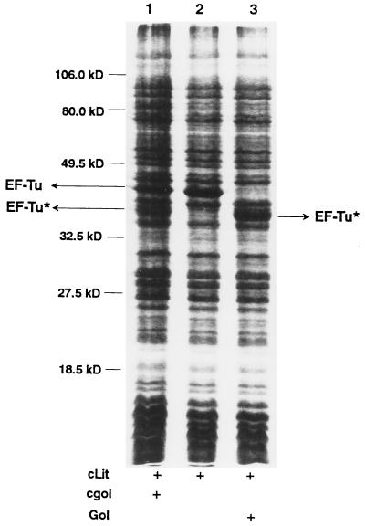
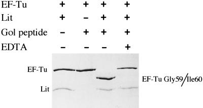
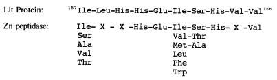
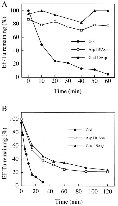
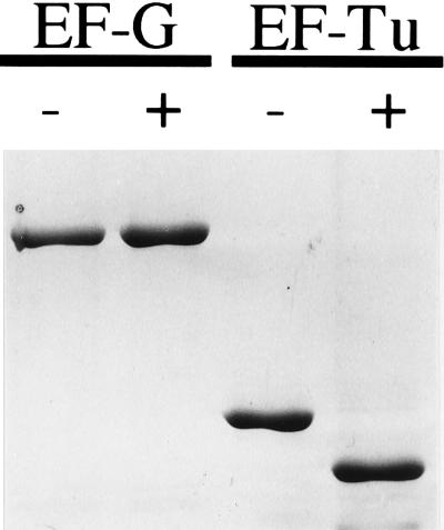
Phage exclusion is a form of programmed cell death in prokaryotes in which death is triggered by infection with phage, a seemingly altruistic response that limits multiplication of the phage and its spread through the population. One of the best-characterized examples of phage exclusion is the exclusion of T-even phages such as T4 by the e14-encoded Lit protein in many Escherichia coli K-12 strains. In this exclusion system, transcription and translation of a short region of the major head coat protein gene late in phage infection activates proteolysis of translation elongation factor Tu (EF-Tu), blocking translation and multiplication of the phage. The cleavage occurs between Gly-59 and Ile-60 in the nucleotide-binding domain. In the present work, we show that a 29-residue synthetic peptide spanning the activating region of the major head coat protein can activate the cleavage of GDP-bound EF-Tu in a purified system containing only purified EF-Tu and purified Lit protein. Lit behaves as a bona fide enzyme in this system, cleaving EF-Tu to completion when present at substoichiometric amounts. Two mutant peptides with amino acid changes that reduce the activation of cleavage of EF-Tu in vivo were also greatly reduced in their ability to activate EF-Tu cleavage in vitro but were still able to activate cleavage at a high concentration. Elongation factor G, which has the same sequence at the cleavage site and a nucleotide-binding domain similar to EF-Tu, was not cleaved by this system, and neither was heat-inactivated EF-Tu, suggesting that the structural context of the cleavage site may be important for specificity. This system apparently represents an activation mechanism for proteolysis that targets one of nature's most evolutionarily conserved proteins for site-specific cleavage.


Figures

Figure 1
Figure 1
Activation of cleavage of EF-Tu in a crude cell lysate by Gol peptide. Two cell extracts, one containing Lit protein and one containing the expressed gol region of T4 phage, were prepared, incubated, and processed as described in Materials and Methods. A Coomassie blue-stained 12% SDS-polyacrylamide gel is shown. Lanes: 1, Gol and Lit-containing extracts mixed before incubation; 2, Lit-containing extract incubated alone; 3, synthetic Gol peptide added to Lit-containing extract before incubation. The positions of EF-Tu and one of the cleavage fragments of EF-Tu (EF-Tu*) are indicated by arrows.
Figure 2
Figure 2
Sequence of the 29-residue synthetic Gol peptide from the major capsid protein of T4 phage. Amino acid changes from the five original mutations identified in the T4 gol region are shown below the sequence, and those chosen for use in this study are underlined. The numbers refer to the position of the amino acid in the gene 23 protein before cleavage during head formation.
Figure 3
Figure 3
EF-Tu can be site-specifically cleaved in a purified three-component system. Purified His-tagged EF-Tu (2 μM) was incubated in the presence or absence of Lit (1 μM) and the synthetic gol peptide (5 μM) in 50 mM Tris⋅HCl, pH 8.0 containing 10 mM 2-mercaptoethanol, 5 mM MgCl2, 10 μM GDP, and 30 μM sarkosyl at 37°C for 1 hr. Samples were then boiled in SDS sample buffer, resolved on a 12% SDS-polyacrylamide gel, and stained with Coomassie blue. Lanes: 1, no Gol peptide; 2, no Lit protein; 3, all three components; 4, all three components but with EDTA added to 10 mM before incubation.
Figure 4
Figure 4
A possible zinc metalloprotease motif in the Lit protein. The consensus histidine residues are putative metal ligation sites. Where there is some variation, the amino acids at those positions are shown. “X” means no apparent consensus at that position. The motif was found by using the GCG wisconsin motifs software program.
Figure 5
Figure 5
Enzymatic activity of Lit and the effect of gol mutations on EF-Tu cleavage. Time course of EF-Tu cleavage by Lit in the presence of wild-type and gol mutant peptides at 2 μM (A) and 200 μM (B) peptide. Incubations were as described in the legend to Fig. 3 and the Materials and Methods section except that the Lit concentration in both sets of data (0.2 μM) was 10% that of substrate EF-Tu. The data are shown as the percentage of starting EF-Tu as determined by laser densitometry of Coomassie blue-stained SDS gels.
Figure 6
Figure 6
EF-G is not a substrate for Lit-mediated cleavage. Purified EF-G and EF-Tu (5 μM) were each incubated with Lit protein with or without Gol peptide (both 5 μM) at 37°C for 1 hr as described in the Materials and Methods. Samples were subjected to SDS/PAGE (10%) and stained with Coomassie blue. The Lit protein has run off the gel so it is not visible.

Comment in

References

    1. Snyder L. Mol Microbiol. 1995;15:415–420. - PubMed
    1. Yarmolinsky M. Science. 1995;267:836–837. - PubMed
    1. Kao C, Snyder L. J Bacteriol. 1988;170:2056–2062. - PMC - PubMed
    1. Hill C W, Gray J A, Brody H. J Bacteriol. 1989;171:4083–4084. - PMC - PubMed
    1. Bergsland K J, Kao C, Yu Y-T N, Gulati R, Snyder L. J Mol Biol. 1990;213:477–494. - PubMed

Publication types

MeSH terms

LinkOut - more resources