. 1998 Mar 17;95(6):2926-31.
doi: 10.1073/pnas.95.6.2926.

Long QT and ventricular arrhythmias in transgenic mice expressing the N terminus and first transmembrane segment of a voltage-gated potassium channel

Affiliations

Long QT and ventricular arrhythmias in transgenic mice expressing the N terminus and first transmembrane segment of a voltage-gated potassium channel

B London et al. Proc Natl Acad Sci U S A. .

Abstract

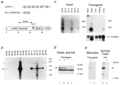
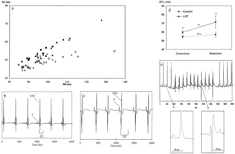
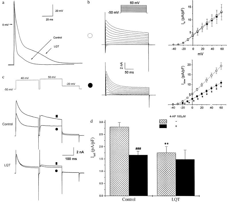
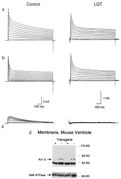
Voltage-gated potassium channels control cardiac repolarization, and mutations of K+ channel genes recently have been shown to cause arrhythmias and sudden death in families with the congenital long QT syndrome. The precise mechanism by which the mutations lead to QT prolongation and arrhythmias is uncertain, however. We have shown previously that an N-terminal fragment including the first transmembrane segment of the rat delayed rectifier K+ channel Kv1.1 (Kv1.1N206Tag) coassembles with other K+ channels of the Kv1 subfamily in vitro, inhibits the currents encoded by Kv1.5 in a dominant-negative manner when coexpressed in Xenopus oocytes, and traps Kv1.5 polypeptide in the endoplasmic reticulum of GH3 cells. Here we report that transgenic mice overexpressing Kv1.1N206Tag in the heart have a prolonged QT interval and ventricular tachycardia. Cardiac myocytes from these mice have action potential prolongation caused by a significant reduction in the density of a rapidly activating, slowly inactivating, 4-aminopyridine sensitive outward K+ current. These changes correlate with a marked decrease in the level of Kv1.5 polypeptide. Thus, overexpression of a truncated K+ channel in the heart alters native K+ channel expression and has profound effects on cardiac excitability.


Figures

Figure 1
Figure 1
Expression of the N-terminal fragment of rat Kv1.1 in the hearts of transgenic mice. (a) Schematic representation of rat brain potassium channel Kv1.1 (Top), the N-terminal fragment Kv1.1N206Tag (Middle), and the transgenic construct (Bottom). HA represents the HA epitope added to the N-terminal fragment of Kv1.1. The α-myosin heavy chain contained ≈1 KB of the promoter followed by exon 1, the first intron, and part of exon 2 (5′ untranslated). N206 represents the N-terminal fragment Kv1.1N206Tag and PA represents the polyadenylation signal. (b) Genomic Southern blot of tail DNA isolated from 16 Fo mice, digested with BamHI, and probed with the fragment of the α-myosin heavy chain denoted in a. The native mouse α-myosin heavy chain gene (∗) and the transgene (arrow) are indicated. Note that each of the seven transgenic founders have multiple copies of the transgene. (c) Northern blot analysis of 2 μg of poly(A)+ RNA (PolyATract, Promega) from the hearts of F1 mice from the lines indicated (Left) and of 15 μg of total RNA (RNEasy, Qiagen) from the indicated organs of an F1 mouse of the B102 line (Right), probed with the N-terminal fragment of Kv1.1. Relative loading of RNA was confirmed by reprobing with a 700-bp fragment of the β-actin gene. (d) Western blot analysis of the steady-state levels of Kv1.1N206Tag polypeptide present in the membrane fraction of LQT hearts (lanes 1–2) and control hearts (lanes 3–4) using a rabbit polyclonal antibody directed against the HA epitope (Santa Cruz Biotechnology). The arrow indicates the position of Kv1.1N206Tag. (e) Immunoprecipitation analysis of Kv1.1N206Tag expressed in primary culture of adult mouse rod shape cardiocytes derived from two LQT mice (lane 1), control cardiocytes (lane 2), and GH3 cells stably transfected with Kv1.1N206Tag (GHT69) (lane 3). In lane 4, the immunoprecipitation was blocked with 90 nmols of HA peptide. Total cell lysates were immunoprecipitated with anti-HA antibody (12CA5). The pellets were analyzed on SDS/PAGE. The arrow indicates the position of Kv1.1N206Tag.
Figure 2
Figure 2
Analysis of the rhythm and QT intervals of ECGs obtained from LQT and control mice. (a) The relationships between the observed QT interval and the RR intervals in the conscious LQT mice (⧫, •, and ▴, n = 10) and control mice (▵, ◊, ×, +, n = 10). Each point represents an average of 72 measurements of the QT and RR intervals (every 20 min) per 24 hr. Each measurement is an average of a 4-sec screen. Each mouse was monitored continuously for 3 days and is therefore represented by three points. (b) The ECG of an anesthetized 3-month-old control mouse (50 mg/kg ketamine and 10 mg/kg xylazine) recorded by the transmitter. The observed heart rate was 180 beats per minute (bpm), and the observed QT interval was 101 ms. (c) The ECG of an anesthetized matched LQT mouse (50 mg/kg ketamine and 10 mg/kg xylazine) recorded by the transmitter. The observed heart rate was 180 bpm, and the observed QT interval was 179 ms. (d) Sensitivity of the QTc interval of LQT mice to ketamine. The average QTc interval of anesthetized and conscious LQT and control mice (average of 2 hr after 50 mg/kg ketamine administration). The ketamine-induced prolongation of the QTc interval in LQT mice was significantly greater than that of control. (e) Surface ECG recording of an 11-beat run of ventricular tachycardia in a conscious freely moving LQT mouse. Note the atrio-ventricular dissociation. The enlarged QRS complexes demonstrate that the individual QRS complexes in the nonsustained ventricular tachycardia differ substantially from the sinus QRS complexes. The sinus heart rate is about 530 bpm (RR interval of 113 ms). The rate of the ventricular tachycardia is 570 bpm (RR interval of 105 ms).
Figure 3
Figure 3
Characterization of AP and the outward potassium currents in LQT cardiocytes. (a) Transmembrane AP recorded from a representative control and a LQT mouse cardiocyte. APs were elicited by suprathreshold current injected through the electrode at 0.5 Hz. For better superimposition, stimulatory artifacts resulting from current injection were minimized off-line. (b) Voltage-dependent K+ current traces and current-voltage relationships in control (∗; n = 24) and LQT mouse ventricular myocytes (•; n = 16). Traces from representative cells are shown (Left). Current density was obtained by normalizing the current amplitude to cell capacitance. The currents were elicited by 10 mV step depolarizations between −40 to +60 mV from −50 mV holding potential. Ito was defined as the difference between the peak outward current and the current level at the end of the 250-ms pulse. The current remaining after 250 ms was significantly lower in LQT myocytes (P < 0.05). (c) Effects of 50 μM 4-AP on the outward currents of a representative ventricular myocyte derived from either a control or a matched LQT mouse. The voltage protocol is shown (Top). ▪ and • indicate the current traces before and after application of 4-AP respectively. (d) Summary of the effects of 100 μM 4-AP on Itail of control and LQT myocytes. Itail was defined as the difference between the peak and the current at the end of −20-mV pulse. The density of Itail was significantly larger in control cardiocytes compared with LQT (∗∗, P < 0.01). 4-AP abolished over 40% of Itail in control cardiocytes (###, P < 0.001), but had no significant effect in LQT cardiocytes.
Figure 4
Figure 4
Effect of 4-AP on outward K+ currents. (ac) Examples of outward K+ currents recorded from mouse cardiocytes derived from control and LQT mice before (a) and after (b) application of 50 μM 4-AP. c illustrates the 4-AP-sensitive components obtained by the subtraction of the currents in b from the currents in aa. A series of prolonged (1,000 ms) test pulses were applied to elicit the outward currents. (d) Western blot analysis of the steady-state levels of Kv1.5 in membrane preparations from the hearts of LQT mice and nontransgenic littermate controls, using a rabbit polyclonal antibody directed against Kv1.5 (Upstate Biotechnology). A gel loaded with the same amount of membrane extract (not boiled) and incubated with an antibody directed against the α subunit of the rat Na-K ATPase demonstrated equal loading of membrane extracts, as did another gel stained with Coumassie blue (data not shown). Similar results were obtained for three transgenic and three control mice, and the blot was repeated in triplicate.

References

    1. Curran M E, Splawski I, Timothy K W, Vincent G M, Green E D, Keating M T. Cell. 1995;80:795–803. - PubMed
    1. Wang Q, Curran M E, Splawski I, Burn T C, Millholland J M, Van Raay T J, Shen J, Timothy K W, Vincent G M, de Jager T, et al. Nat Genet. 1996;12:17–23. - PubMed
    1. Sanguinetti M C, Jiang C, Curran M E, Keating M T. Cell. 1995;81:299–307. - PubMed
    1. Trudeau M C, Waronhe J M, Ganetsky B, Robertson G A. Science. 1995;269:92–95. - PubMed
    1. Sanguinetti M C, Curran M E, Zou A, Shen J, Spector P S, Atkinson D L, Keating M T. Nature (London) 1996;384:80–83. - PubMed

Publication types

MeSH terms