. 1998 Mar 17;95(6):2944-9.
doi: 10.1073/pnas.95.6.2944.

A large and distinct rotation of the myosin light chain domain occurs upon muscle contraction

Affiliations

A large and distinct rotation of the myosin light chain domain occurs upon muscle contraction

J E Baker et al. Proc Natl Acad Sci U S A. .

Abstract

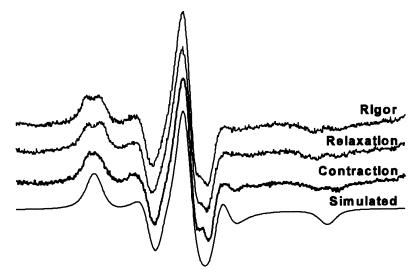
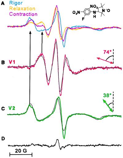
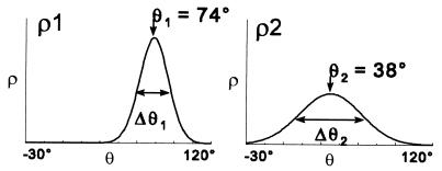
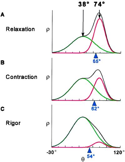
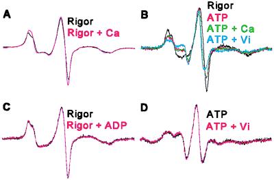
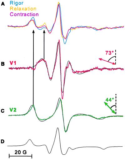
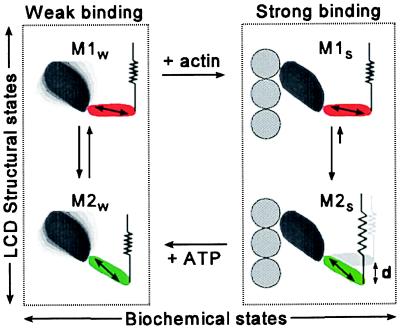
For more than 30 years, the fundamental goal in molecular motility has been to resolve force-generating motor protein structural changes. Although low-resolution structural studies have provided evidence for force-generating myosin rotations upon muscle activation, these studies did not resolve structural states of myosin in contracting muscle. Using electron paramagnetic resonance, we observed two distinct orientations of a spin label attached specifically to a single site on the light chain domain of myosin in relaxed scallop muscle fibers. The two probe orientations, separated by a 36 degrees +/- 5 degrees axial rotation, did not change upon muscle activation, but the distribution between them changed substantially, indicating that a fraction (17% +/- 2%) of myosin heads undergoes a large (at least 30 degrees) axial rotation of the myosin light chain domain upon force generation and muscle contraction. The resulting model helps explain why this observation has remained so elusive and provides insight into the mechanisms by which motor protein structural transitions drive molecular motility.


Figures

Figure 1
Figure 1
EPR spectra of oriented fibers. Spectra shown (from top, down) are fibers in rigor, relaxation, and contraction. The significant difference between the spectra of fibers oriented parallel (black) and perpendicular (gray) to the magnetic field shows that the probes are well ordered in all three physiological states.
Figure 2
Figure 2
EPR spectra of randomly oriented fibers. Spectra shown (from top, down) are fibers in rigor, relaxation, contraction, and simulation of the best fit to the rigor experimental spectrum. The simulated spectrum has a Lorentzian line width of 2.2 G, Gaussian line width of 2.8 G, anisotropic T values Tzz = 29.9 G, Tyy = 9.8 G, and Txx = 8.3 G, and g values gxx = 2.0072, gyy = 2.0068, and gzz = 2.0035.
Figure 3
Figure 3
Two resolved orientational components derived from EPR spectra of spin-labeled scallop muscle fibers. (A) Spectra overlaid in rigor (cyan), relaxation (orange), and contraction (magenta). (B and C) Resolved spectral components V1 (red) and V2 (green) were obtained by subtraction of rigor and relaxation spectra, yielding unique endpoints (9). Each of the black spectra in B and C is the best-fit simulation (34) to a single, oriented population of the probe’s principal axis (double-headed arrow) with respect to the fiber axis (vertical, dashed line). The residual spectrum (D) shows the small difference between the contraction spectrum and a linear combination of V1 and V2 (Eq. 1, x1 = 0.33, x2 = 0.67).
Figure 4
Figure 4
Gaussian orientational distribution for each component determined by fitting EPR spectral components V1 and V2 (Fig. 3) to simulated spectra corresponding to ρi(θ) = exp − {(ln2)[(θ − θi)/(Δθi/2)]2}/∫(ρi(θ)sinθdθ).
Figure 5
Figure 5
The overall angular distribution ρ(θ) (Eq. 1, plotted in black), of spin-labeled LC domain in scallop fibers for different physiological states, showing contributions from the two oriented components, ρ1 (red) and ρ2 (green), as determined from the EPR spectra in Fig. 2. The average value of θ for the overall distribution, 〈θ〉 = ∫θsin(θ)ρ(θ)dθ, is indicated in blue. (A) Relaxation: ρ1 and ρ2 are both populated. (B) Contraction: the distribution shifts toward ρ2. (C) Rigor: ρ2 is predominant.
Figure 6
Figure 6
Controls. (A) Addition of calcium to rigor had no significant effect on the spectrum. (B) Partially extracted fibers, which are activated by ATP with or without calcium, show essentially the same spectra as unextracted fibers that have been Ca-activated. On addition of vanadate, the overall distribution shifts toward that of relaxation. (C) Addition of ADP to rigor and (D) addition of vanadate to relaxation had little effect on the original spectrum.
Figure 7
Figure 7
EPR spectra of spin-labeled rabbit muscle fibers. (A) Spectra overlaid in rigor (cyan), relaxation (orange), and contraction (magenta). (B and C) Orientational components V1 (red) and V2 (green) were obtained as in Fig. 2, with the additional subtraction of up to 40% of a powder spectrum (D). Black spectra in B and C are the best-fit simulations of Gaussian distributions centered at 73 ± 5° and 44 ± 5° with full width at half-maximum of 24 ± 5° and 40 ± 5°, respectively.
Figure 8
Figure 8
A myosin crossbridge model. Force is generated upon the transition from weak (Left) to strong (Right) actin binding, which is coupled to a change in the distribution between two myosin head structures, M1 and M2, having LC domain orientations that differ by at least 36°. The stretched spring (Right Lower) indicates that force has been produced and does not necessarily correspond to a specific structural feature.

Comment in

  • New angle on myosin.
    Cooke R. Cooke R. Proc Natl Acad Sci U S A. 1998 Mar 17;95(6):2720-2. doi: 10.1073/pnas.95.6.2720. Proc Natl Acad Sci U S A. 1998. PMID: 9501153 Free PMC article. No abstract available.

References

    1. Huxley H E. Science. 1969;164:1356–1366. - PubMed
    1. Huxley A F. J Physiol (London) 1974;243:1–42. - PMC - PubMed
    1. Lymn R W, Taylor E W. Biochemistry. 1971;10:4617–4624. - PubMed
    1. Huxley H E, Simmons R M, Faruqi A R, Kress M, Bordas J, Koch M H J. J Mol Biol. 1983;169:469–506. - PubMed
    1. Irving M. J Physiol (London) 1993;472:127–156. - PMC - PubMed

Publication types

LinkOut - more resources