. 1998 Mar 17;95(6):2979-84.
doi: 10.1073/pnas.95.6.2979.

HL-1 cells: a cardiac muscle cell line that contracts and retains phenotypic characteristics of the adult cardiomyocyte

Affiliations

HL-1 cells: a cardiac muscle cell line that contracts and retains phenotypic characteristics of the adult cardiomyocyte

W C Claycomb et al. Proc Natl Acad Sci U S A. .

Abstract

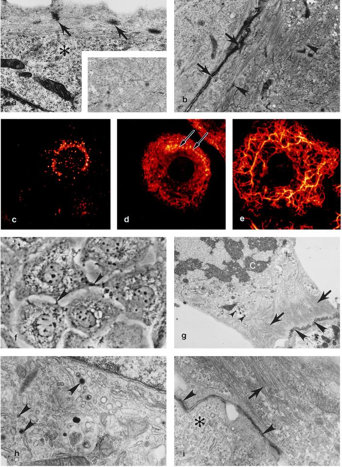
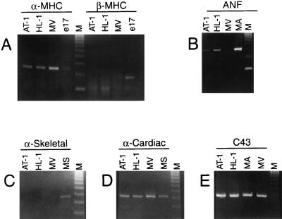
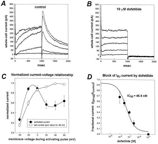
We have derived a cardiac muscle cell line, designated HL-1, from the AT-1 mouse atrial cardiomyocyte tumor lineage. HL-1 cells can be serially passaged, yet they maintain the ability to contract and retain differentiated cardiac morphological, biochemical, and electrophysiological properties. Ultrastructural characteristics typical of embryonic atrial cardiac muscle cells were found consistently in the cultured HL-1 cells. Reverse transcriptase-PCR-based analyses confirmed a pattern of gene expression similar to that of adult atrial myocytes, including expression of alpha-cardiac myosin heavy chain, alpha-cardiac actin, and connexin43. They also express the gene for atrial natriuretic factor. Immunohistochemical staining of the HL-1 cells indicated that the distribution of the cardiac-specific markers desmin, sarcomeric myosin, and atrial natriuretic factor was similar to that of cultured atrial cardiomyocytes. A delayed rectifier potassium current (IKr) was the most prominent outward current in HL-1 cells. The activating currents displayed inward rectification and deactivating current tails were voltage-dependent, saturated at >>+20 mV, and were highly sensitive to dofetilide (IC50 of 46.9 nM). Specific binding of [3H]dofetilide was saturable and fit a one-site binding isotherm with a Kd of 140 +/- 60 nM and a Bmax of 118 fmol per 10(5) cells. HL-1 cells represent a cardiac myocyte cell line that can be repeatedly passaged and yet maintain a cardiac-specific phenotype.


Figures

Figure 1
Figure 1
Expression of a cardiac-specific phenotype at multiple passages in the HL-1 cardiomyocyte cell line. (a) A 10th-passage HL-1 cell demonstrating subsarcolemmal Z densities (arrows) typical of normal myofibrillogenesis in cultured and in vivo cardiomyocytes, glycogen (∗). (×12,500.) (Inset) High-magnification view demonstrating typical cardiac-specific myofibril banding. (×20,000.) (b) Two 10th-passage HL-1 cells containing myofibrils at various stages of sarcomerogenesis (arrowheads) are attached via an immature intercalated disc (arrows). (×9,670.) (c) Immunofluorescent localization of ANF expression in a passage 20 HL-1 cell. (d) Myosin is localized to scattered filaments in the cytoplasm and to thin reorganizing myofibrils located along the peripheral cytoplasm (arrows) in a passage 20 HL-1 cell. (e) In a passage 20 HL-1 cell desmin is expressed as reticulated cytoplasmic rings of intermediate filaments extending into lamellapodia. (c–e) False color, indirect immunofluorescence confocal laser scanning microscopy images. (×1,650.) (f) Passage 34 HL-1 cells demonstrating centrally located mononucleation and intercellular junctional contacts (arrowheads); phase-contrast, ×700. (g) At passage 34, dividing HL-1 cells behave like typical mitotic cardiomyocytes in that they retain peripheral myofibrils (arrows), contain atrial granules (small arrowheads,) and are anchored to adjacent cardiomyocytes by intercalated discs (arrowheads). C, chromosomes. (×8,000.) (h) At passage 86, an active Golgi and atrial-specific granules are present (arrowheads). (×25,000.) (i) A passage 86 HL-1 cell containing organized myofibrils (arrow), an intercalated disc (arrowheads), and areas occupied by free ribosomes (∗). (×15,000.) (a, b, and g–i) Transmission electron micrographs.
Figure 2
Figure 2
Reverse transcriptase–PCR-based analyses of gene expression in cultured HL-1 cells. (A) α-MHC and β-MHC, (B) ANF, (C) α-skeletal actin (α-skeletal), (D) α-cardiac actin (α-cardiac), and (E) connexin43 (C43) in controls and cultured HL-1 cells at passage 86. Total RNA was isolated from AT-1 cells (AT-1), HL-1 cells (HL-1), ventricular cardiac muscle tissue from 17 day embryonic rat (e17), atrial muscle tissue from adult mouse (MA), ventricular cardiac muscle tissue from adult mouse (MV), and skeletal muscle tissue from adult mouse (MS). DNA molecular weight markers [φX174 RF DNA, HaeIII cut] (M).
Figure 3
Figure 3
Characteristics of IKr-like current recorded from HL-1 cells. (A) Whole-cell current traces recorded from an HL-1 cell. Voltage pulses (1-sec duration) were applied from a holding potential of −50 mV to test potentials between −40 mV to +40 mV in 20-mV increments. The membrane potential was returned to −50 mV after each depolarizing pulse to better resolve outward tail currents. (B) Whole-cell currents in the presence of 10 μM of dofetilide (same cell as in A). Dofetilide abolished the time-dependent component of the activating current and the deactivating tail currents. Complete blockade of the time-dependent component of the activating outward current revealed a residual time-independent outward current in most HL-1 cells. (C) Current-voltage relationship for the time-dependent activating outward current and the deactivating tail current. Plotted are the normalized magnitudes of whole-cell currents recorded at the end of each 1-sec depolarizing pulse (•) and tail currents (○) versus membrane potential. Currents were normalized as a fraction of peak current magnitude. Values are means ± SEM (n = 10 cells). (D) Concentration-response relationship for block of IKr tail current by dofetilide. IC50 was 46.9 nM (n = 4).

References

    1. Field L J. Science. 1988;239:1029–1033. - PubMed
    1. Steinhelper M E, Lanson N A, Jr, Dresdner K P, Delcarpio J B, Wit A L, Claycomb W C, Field L J. Am J Physiol. 1990;259:H1826–H1834. - PubMed
    1. Delcarpio J B, Lanson N A, Jr, Field L J, Claycomb W C. Circ Res. 1991;69:1591–1600. - PubMed
    1. Lanson N A, Jr, Glembotski C C, Steinhelper M E, Field L J, Claycomb W C. Circulation. 1992;85:1835–1841. - PubMed
    1. Borisov A B, Claycomb W C. Ann NY Acad Sci. 1995;752:80–91. - PubMed

Publication types