

Linkage analysis and comparative mapping of canine progressive rod-cone degeneration (prcd) establishes potential locus homology with retinitis pigmentosa (RP17) in humans
- PMID: 9501213
- PMCID: PMC19692
- DOI: 10.1073/pnas.95.6.3048
Linkage analysis and comparative mapping of canine progressive rod-cone degeneration (prcd) establishes potential locus homology with retinitis pigmentosa (RP17) in humans
Abstract
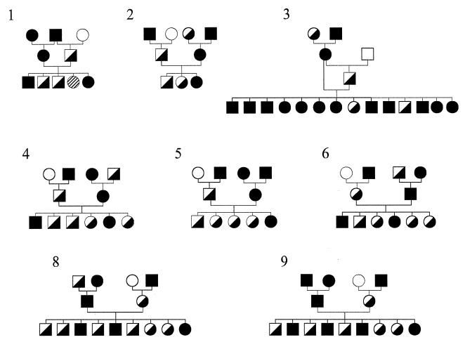
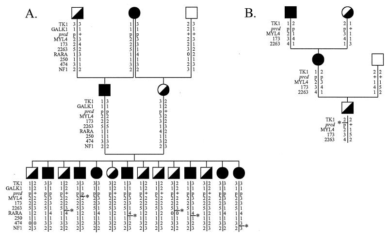
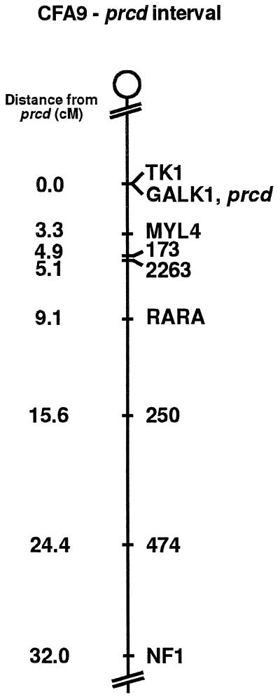
Progressive rod-cone degeneration (prcd) is the most widespread hereditary retinal disease leading to blindness in dogs and phenotypically is the canine counterpart of retinitis pigmentosa (RP) in humans. In previous efforts to identify the genetic locus for prcd, the canine homologs for many of the genes causally associated with RP in humans, such as RHO, PDE6B, and RDS/peripherin, have been excluded. In parallel with a recent undertaking to establish a framework map of the canine genome, multiple prcd-informative pedigrees have been typed with a panel of more than 100 anchor loci and microsatellite-based markers. Identification of a linkage group flanking prcd ([TK1, GALK1, prcd]-[MYL4, C09.173, C09.2263]-RARA-C09.250-C09.474-NF1) localizes prcd close to the centromeric end of canine chromosome 9 (CFA9), and excludes RARA as a candidate gene. The conserved synteny of this region of CFA9 and distal human chromosome 17q establishes the potential locus homology of prcd in the dog with RP17, a human retinitis pigmentosa locus for which no gene has yet been identified. Assignment of the prcd disease locus to an identified canine autosome represents a powerful application of the developing canine linkage map in medical genetics. The usefulness of this approach is further demonstrated by identification of the correspondence of the prcd interval to homologous human and mouse chromosomal regions. The rapid progress that is now occurring in the field of canine genetics will expedite the identification of the genes underlying many of the inherited traits and diseases that make the dog a unique asset for the study of mammalian traits.
Figures
References
-
- Little C C. The Inheritance of Coat Color in Dogs. Ithaca, NY: Comstock; 1957.
-
- Clark R D, Stainer J R, editors. Medical and Genetic Aspects of Purebred Dogs. Edwardsville, KS: Veterinary Medicine Publishing; 1983.
-
- Patterson D F, Haskins M E, Jezyk P F. Adv Hum Genet. 1982;12:263–339. - PubMed
-
- Patterson D F, Haskins M E, Jezyk P F, Giger U, Meyers-Wallen V N, Aguirre G, Fyfe J C, Wolfe J H. J Am Vet Med Assoc. 1988;193:1131–1144. - PubMed
-
- Bech-Nielsen S, Haskins M E, Reif J S, Brodey R S, Patterson D F, Spielman R. J Natl Cancer Inst. 1978;60:349–353. - PubMed
Publication types
MeSH terms
Substances
Associated data
- Actions
Grants and funding
LinkOut - more resources
Full Text Sources
Other Literature Sources
Research Materials
Miscellaneous
