

Phosphatidylserine exposure and red cell viability in red cell aging and in hemolytic anemia
- PMID: 9501218
- PMCID: PMC19697
- DOI: 10.1073/pnas.95.6.3077
Phosphatidylserine exposure and red cell viability in red cell aging and in hemolytic anemia
Abstract
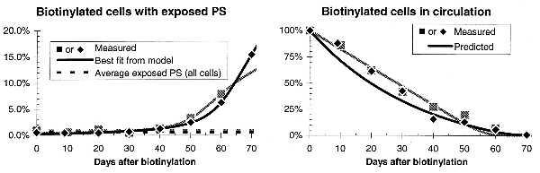
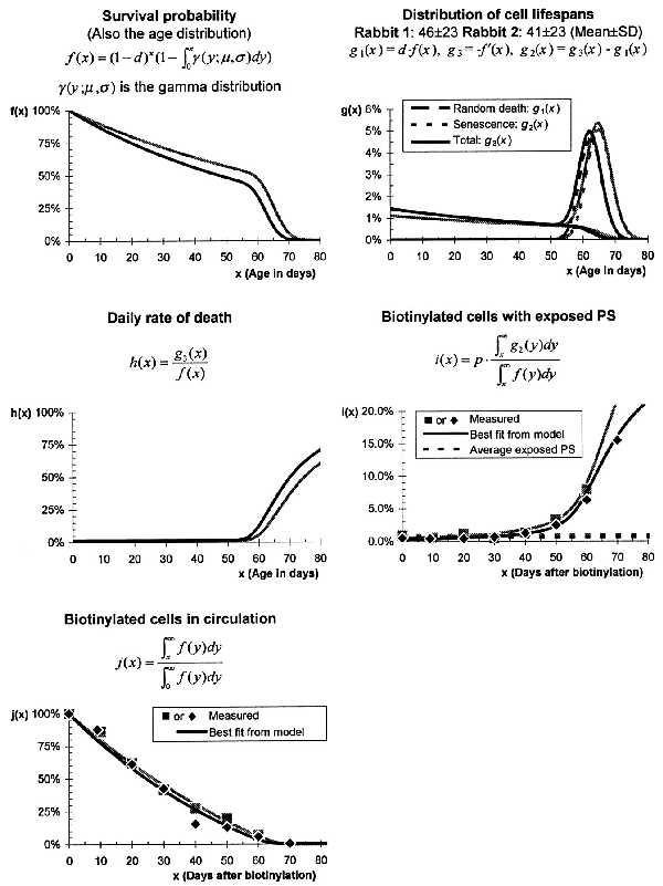
Phosphatidylserine (PS) normally localizes to the inner leaflet of cell membranes but becomes exposed in abnormal or apoptotic cells, signaling macrophages to ingest them. Along similar lines, it seemed possible that the removal of red cells from circulation because of normal aging or in hemolytic anemias might be triggered by PS exposure. To investigate the role of PS exposure in normal red cell aging, we used N-hydroxysuccinimide-biotin to tag rabbit red cells in vivo, then used phycoerythrin-streptavidin to label the biotinylated cells, and annexin V-fluorescein isothiocyanate (FITC) to detect the exposed PS. Flow cytometric analysis of these cells drawn at 10-day intervals up to 70 days after biotinylation indicated that older, biotinylated cells expose more PS. Furthermore, our data match a simple model of red cell senescence that assumes both an age-dependent destruction of senescent red cells preceded by several hours of PS exposure and a random destruction of red cells without PS exposure. By using this model, we demonstrated that the exposure of PS parallels the rate at which biotinylated red cells are removed from circulation. On the other hand, using an annexin V-FITC label and flow cytometry demonstrates that exposed PS does not cause the reduced red cell life span of patients with hemolytic anemia, with the possible exception of those with unstable hemoglobins or sickle cell anemia. Thus, in some cases PS exposure on the cell surface may signal the removal of red cells from circulation, but in other cases some other signal must trigger the sequestration of cells.
Figures

References
-
- Diaz C, Schroit A J. J Membr Biol. 1996;151:1–9. - PubMed
-
- Yaffe M P, Kennedy E P. Biochemistry. 1983;22:1497–1507. - PubMed
-
- Savill J, Fadok V, Henson P, Haslett C. Immunol Today. 1993;3:131–136. - PubMed
-
- Fadok V A, Laszlo D J, Noble P W, Weinstein L, Riches D W, Henson P M. J Immunol. 1993;151:4274–4285. - PubMed
-
- Fadok V A, Voelker D R, Campbell P A, Cohen J J, Bratton D L, Henson P M. J Immunol. 1992;148:2207–2216. - PubMed
Publication types
MeSH terms
Substances
Grants and funding
LinkOut - more resources
Full Text Sources
Other Literature Sources
