

Functional anatomy of musical processing in listeners with absolute pitch and relative pitch
- PMID: 9501235
- PMCID: PMC19714
- DOI: 10.1073/pnas.95.6.3172
Functional anatomy of musical processing in listeners with absolute pitch and relative pitch
Abstract
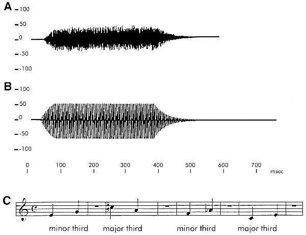
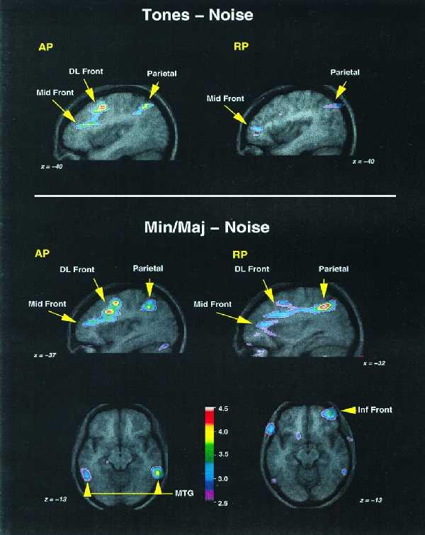
We used both structural and functional brain imaging techniques to investigate the neural basis of absolute pitch (AP), a specialized skill present in some musicians. By using positron emission tomography, we measured cerebral blood flow during the presentation of musical tones to AP possessors and to control musicians without AP. Listening to musical tones resulted in similar patterns of increased cerebral blood flow in auditory cortical areas in both groups, as expected. The AP group also demonstrated activation of the left posterior dorsolateral frontal cortex, an area thought to be related to learning conditional associations. However, a similar pattern of left dorsolateral frontal activity was also observed in non-AP subjects when they made relative pitch judgments of intervals, such as minor or major. Conversely, activity within the right inferior frontal cortex was observed in control but not in AP subjects during the interval-judgment task, suggesting that AP possessors need not access working memory mechanisms in this task. MRI measures of cortical volume indicated a larger left planum temporale in the AP group, which correlated with performance on an pitch-naming task. Our findings suggest that AP may not be associated with a unique pattern of cerebral activity but rather may depend on the recruitment of a specialized network involved in the retrieval and manipulation of verbal-tonal associations.
Figures
References
-
- Ward W D, Burns E M. In: The Psychology of Music. Deutsch D, editor. New York: Academic; 1982. pp. 431–451.
-
- Takeuchi A H, Hulse S H. Psychol Bull. 1993;113:345–361. - PubMed
-
- Zatorre R J. Cortex. 1989;25:567–580. - PubMed
-
- Schlaug G, Jäncke L, Huang Y, Steinmetz H. Science. 1995;267:699–701. - PubMed
-
- Galaburda A, Sanides F. J Comp Neurol. 1980;190:597–610. - PubMed
Publication types
MeSH terms
LinkOut - more resources
Full Text Sources
