. 1998 Mar 17;95(6):3281-6.
doi: 10.1073/pnas.95.6.3281.

Differential colocalization of estrogen receptor beta (ERbeta) with oxytocin and vasopressin in the paraventricular and supraoptic nuclei of the female rat brain: an immunocytochemical study

Affiliations

Differential colocalization of estrogen receptor beta (ERbeta) with oxytocin and vasopressin in the paraventricular and supraoptic nuclei of the female rat brain: an immunocytochemical study

S E Alves et al. Proc Natl Acad Sci U S A. .

Abstract

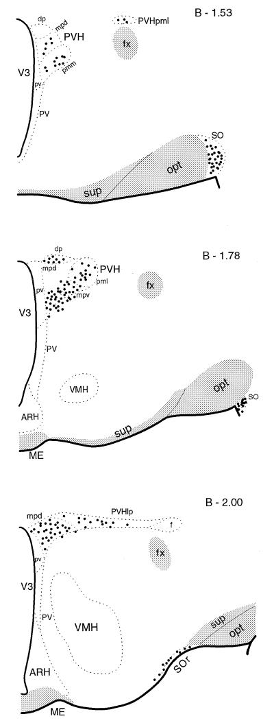
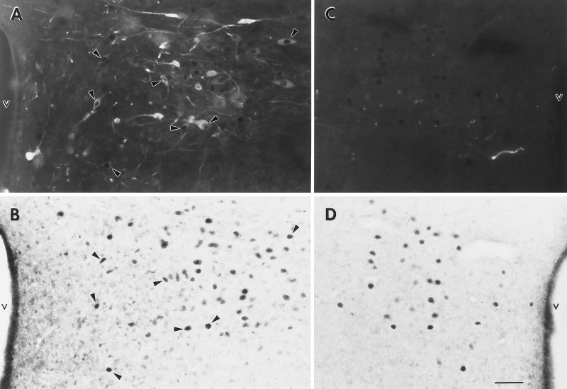
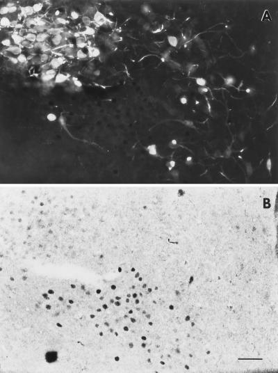
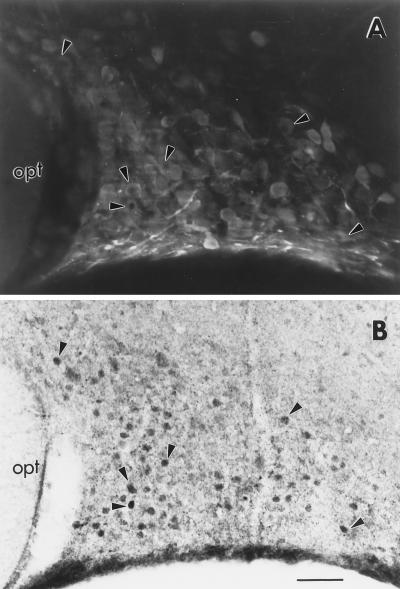
Evidence exists for the localization of the newly identified estrogen receptor beta (ERbeta) within the rat paraventricular nucleus (PVN) and supraoptic nucleus (SON), regions which lack ERalpha. Presently, we investigate whether ERbeta-like-immunoreactivity (-ir) is found within cells of several major neuropeptide systems of these regions. Young adult Sprague-Dawley rats were ovariectomized (OVX), and 1 week later half of the animals received estradiol-17beta (E). Dual-label immunocytochemistry was performed on adjacent sections by using an ERbeta antibody, followed by an antibody to either oxytocin (OT), arginine-vasopressin (AVP), or corticotropin releasing hormone. Nuclear ERbeta-ir was identified within SON and retrochiasmatic SON, and in specific PVN subnuclei: medial parvicellular part, ventral and dorsal zones, dorsal and lateral parvicellular parts, and in the posterior magnocellular part, medial and lateral zones. However, the ERbeta-ir within magnocellular areas was noticeably less intense. OT-/ERbeta-ir colocalization was confirmed in neurons of the parvicellular subnuclei, in both OVX and OVX+E brains ( approximately 50% of OT and 25% of ERbeta-labeled cells between bregma -1.78 and -2.00). In contrast, few PVN parvicellular neurons contained both AVP- and ERbeta-ir. As well, very little overlap was observed in the distribution of cells containing corticotropin releasing hormone- or ERbeta-ir. In the SON, most nuclear ERbeta-ir colocalized with AVP-ir, whereas few OT-/ERbeta-ir dual-labeled cells were observed. These findings suggest that estrogen can directly modulate specific OT and AVP systems through an ERbeta-mediated mechanism, in a tissue-specific manner.


Figures

Figure 1
Figure 1
Brain maps are modified from Swanson (19) and represent coronal sections from three levels of the brain measured from bregma (B). Approximate distributions of cells containing nuclear ERβ-ir in the PVN (PVH in the figure) and SON (SO in the figure) of the female rat hypothalamus are illustrated. Each dot represents approximately two labeled cells within a 40-μm section. fx, Fornix; ME, median emminence; opt, optic tract; sup, supraoptic commissures; V3, third ventricle; sudivisions of the PVN: dp, dorsal parvicellular; lp, lateral parvicellular; mpd, medial parvicellular, dorsal zone; mpv, medial parvicellular, ventral zone; pv, periventricular part; pmm, posterior magnocellular, medial zone; pml, posterior magnocellular, lateral zone; SOr, retrochiasmatic SON; ARH, arcuate nucleus; PV, periventricular nucleus; VMH, ventromedial nucleus.
Figure 2
Figure 2
Fluorescent OT-ir (A) and light-field nuclear ERβ-ir (B) in the PVN of a 40-μm-thick section at the level of approximately B −1.90. Arrowheads denote examples of dual-labeled cells (compare arrowheads in A and B). Note the absence of cell soma AVP-ir at a comparable level (C), where numerous cells containing nuclear ERβ-ir are seen (D). Photomicrographs were taken at ×200 under a Nikon light microscope and a 35 mm camera by using Kodak technical pan film. v, Third ventricle. (Bar = 40 μm.)
Figure 3
Figure 3
Fluorescent AVP-ir (A) and light-field nuclear ERβ-ir (B) in the PVN of a 40-μm section, approximately at B −1.78. Note the very dark, well-defined nuclear ERβ-ir within the lower, central half of panel (B), which corresponds primarily to the medial parvicellular, ventral, and dorsal zones. In contrast, note the very light, yet distinct ERβ-ir in the upper left corner of B, which corresponds to the posterior magnocellular part, lateral zone. Scattered cells containing AVP-ir were found in the parvicellular regions, but only an occasional cell appeared to contain both AVP- and ERβ-ir (none demonstrated here). Most ERβ-ir within the magnocellular area appeared to be located adjacent to, but not in, the AVP-ir magnocellular neurons. Photomicrographs were taken at ×200 under a Nikon light microscope and a 35 mm camera by using Kodak technical pan film. (Bar = 40 μm.)
Figure 4
Figure 4
Fluorescent AVP-ir (A) and light-field nuclear ERβ-ir (B) in the SON of a 40-μm section, at approximately B −1.08. Arrowheads denote examples of dual-labeled cells (compare arrowheads in A and B). Throughout the SON, many of the ERβ-labeled cells also contained AVP-ir. Photomicrographs were taken at ×200 under a Nikon light microscope and a 35 mm camera by using Kodak technical pan film. opt, Optic tract. (Bar = 40 μm.)

References

    1. Kuiper G G J M, Enmark E, Pelto-Huikko M, Nilsson S, Gustafsson J-A. Proc Natl Acad Sci USA. 1996;93:5925–5930. - PMC - PubMed
    1. Mosselman S, Polman J, Dijkema R. FEBS Lett. 1996;392:49–53. - PubMed
    1. Tremblay G B, Tremblay A, Copeland N G, Gilbert D J, Jenkins N, Labrie F, Giguere V. Mol Endocrinol. 1997;11:353–365. - PubMed
    1. Kuiper G G J M, Carlsson B, Grandien K, Enmark E, Haggblad J, Nilsson S, Gustafsson J-A. Endocrinology. 1997;138:863–870. - PubMed
    1. Paech K, Webb P, Kuiper G G J M, Nilsson S, Gustafsson J-A, Kushner P, Scanlan T S. Science. 1997;277:1508–1510. - PubMed

Publication types

MeSH terms