. 1998 Mar;64(3):914-21.
doi: 10.1128/AEM.64.3.914-921.1998.

Biochemical and genetic characterization of an extracellular protease from Pseudomonas fluorescens CY091

Affiliations

Biochemical and genetic characterization of an extracellular protease from Pseudomonas fluorescens CY091

C H Liao et al. Appl Environ Microbiol. 1998 Mar.

Abstract

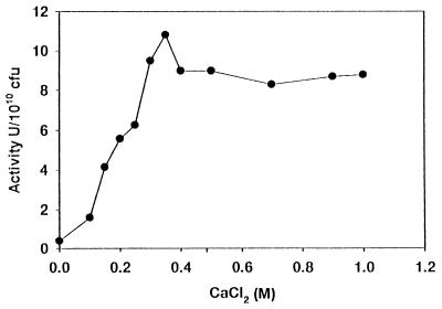
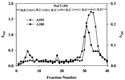
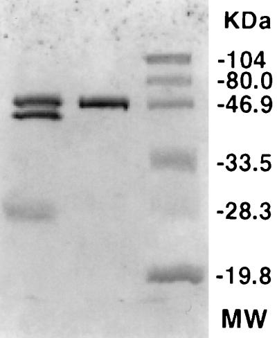
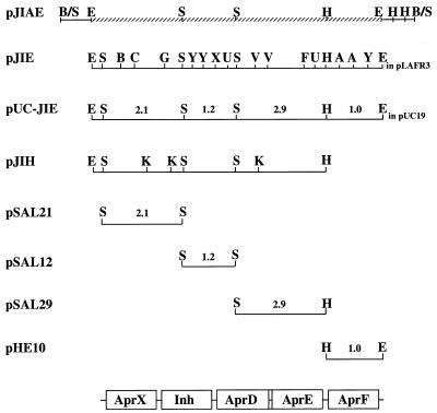
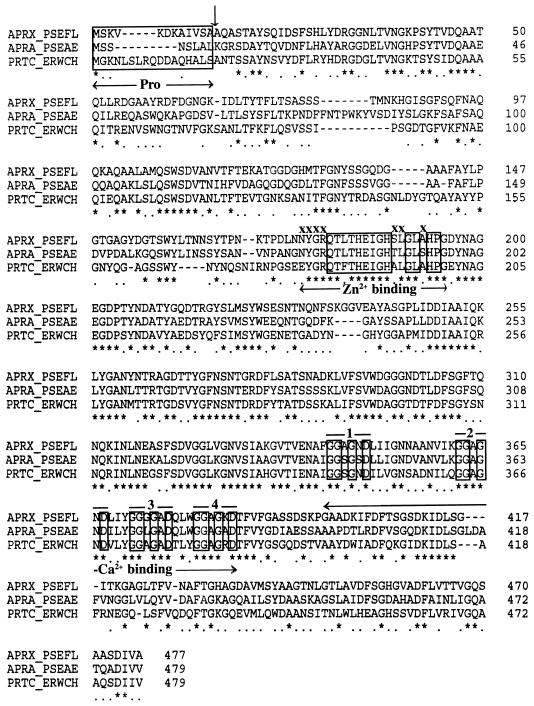
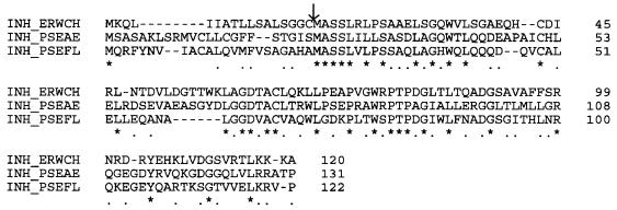
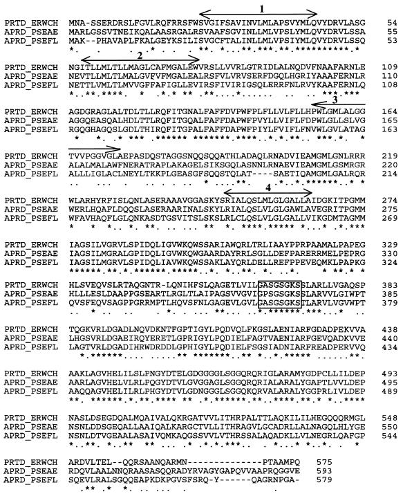
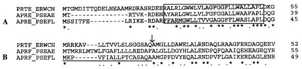
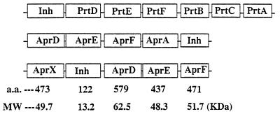
Pseudomonas fluorescens CY091 cultures produce an extracellular protease with an estimated molecular mass of 50 kDa. Production of this enzyme (designated AprX) was observed in media containing CaCl2 or SrCl2 but not in media containing ZnCl2, MgCl2, or MnCl2. The requirement of Ca2+ (or Sr2+) for enzyme production was concentration dependent, and the optimal concentration for production was determined to be 0.35 mM. Following ammonium sulfate precipitation and ion-exchange chromatography, the AprX in the culture supernatant was purified to near electrophoretic homogeneity. Over 20% of the enzyme activity was retained in the AprX sample which had been heated in boiling water for 10 min, indicating that the enzyme is highly resistant to heat inactivation. The enzyme activity was almost completely inhibited in the presence of 1 mM 1,10-phenanthroline, but only 30% of the activity was inhibited in the presence of 1 mM EGTA. The gene encoding AprX was cloned from the genome of P. fluorescens CY091 by isolating cosmid clones capable of restoring the protease production in a nonproteolytic mutant of strain CY091. The genomic region of strain CY091 containing the aprX gene was located within a 7.3-kb DNA fragment. Analysis of the complete nucleotide sequence of this 7.3-kb fragment revealed the presence of a cluster of genes required for the production of extracellular AprX in P. fluorescens and Escherichia coli. The AprX protein showed 50 to 60% identity in amino acid sequence to the related proteases produced by Pseudomonas aeruginosa and Erwinia chrysanthemi. Two conserved sequence domains possibly associated with Ca2+ and Zn2+ binding were identified. Immediately adjacent to the aprX structural gene, a gene (inh) encoding a putative protease inhibitor and three genes (aprD, aprE, and aprF), possibly required for the transport of AprX, were also identified. The organization of the gene cluster involved in the synthesis and secretion of AprX in P. fluorescens CY091 appears to be somewhat different from that previously demonstrated in P. aeruginosa and E. chrysanthemi.


Figures

FIG. 1
FIG. 1
Effects of CaCl2 concentrations on protease production by P. fluorescens CY091 in MS medium (17).
FIG. 2
FIG. 2
Elution profile of the AprX protease of P. fluorescens CY091 from the DEAE-cellulose column. The column was eluted with 50 mM Tris-HCl (pH 8.0) buffer followed by stepwise elution with buffer containing 0.1 to 0.5 M NaCl. The protease activity of each fraction, as indicated by the absorbance at 595 nm (A595), was determined under the conditions described in Materials and Methods. One unit of protease activity is defined as the amount of enzyme which causes an increase of 1 absorbance unit at 595 nm.
FIG. 3
FIG. 3
SDS-polyacrylamide gel electrophoresis of the purified AprX protease sample from P. fluorescens CY091. Molecular mass (MW) standards (in kilodaltons) are shown in the right-hand lane: phosphorylase b, 104; bovine serum albumin, 80; ovalbumin, 46.9; carbonic anhydrase, 33.5; soybean trypsin inhibitor, 28.3; lysozyme, 19.8. Purified AprX and the ammonium sulfate-precipitated sample (50 to 95% saturation) are shown in the middle and left lanes, respectively.
FIG. 4
FIG. 4
Restriction map of P. fluorescens CY091 genomic DNA encoding the aprX protease gene and the aprD, aprE, and aprF genes required for enzyme secretion. The relative positions of these genes and a gene coding for a putative protease inhibitor (Inh) are indicated at the bottom. The length of the fragment in kilobase pairs is indicated above the line. Restriction enzymes: B, BamHI; S, SalI; K, KpnI; H, HindIII; E, EcoRI; C, ClaI; Y, StyI; G, BglII; U, StuI; X, XhoI; F, AflIII; A, AvaI; B/S, BamHI/Sau3A. The hatched area indicates the region where the nucleotide sequence has been determined. pJIE and pUC-JIE are derived from the insertion of the 7.3-kb EcoRI aprX+ fragment into pLAFR3 and pUC19, respectively. pJIH is a deletion subclone of pJIAE.
FIG. 5
FIG. 5
Multiple-sequence alignment of zinc proteases from P. fluorescens CY091 (APRX_PSEFL) (this study), P. aeruginosa PA01 (APRA_PSEAE) (7), and E. chrysanthemi B374 (PRTC_ERWCH) (16).
FIG. 6
FIG. 6
Amino acid sequence alignment of the protease inhibitor (INH) from P. fluorescens CY091 (INH_PSEFL), P. aeruginosa (INH_PSEAE), and E. chrysanthemi (INH_ERWCH). The arrow indicates the predicted cleavage site of the signal peptidase.
FIG. 7
FIG. 7
Multiple-sequence alignment of the AprD proteins from P. fluorescens CY091 (APRD_PSEFL) and P. aeruginosa (APRD_PSEAE) and the PrtD protein from E. chrysanthemi (PRTD_ERWCH). The numbered lines above the sequence indicate the hydrophobic regions. The boxed area indicates the ATP-binding site.
FIG. 8
FIG. 8
Alignment and comparison of the amino-terminal sequences of the AprE (A) and AprF (B) proteins from P. fluorescens CY091 (APRE/F_PSEFL) and P. aeruginosa (APRE/F_PSEAE) and the PrtE/F proteins from E. chrysanthemi (PRTE/F_ERWCH). The boxed area in panel A represents the hydrophobic region, and the underline in panel B represents the putative signal peptide sequence. The arrow indicates the possible peptidase cleavage site.
FIG. 9
FIG. 9
Comparison of the organization of the gene cluster involved in the synthesis and secretion of the AprX protease in P. fluorescens, the AprA protease in P. aeruginosa, and the PrtB, PrtC, and PrtA proteases in E. chrysanthemi. The predicted amino acid residues and molecular mass (MW) of each protein component in the P. fluorescens AprX gene cluster are indicated at the bottom of the figure.

Similar articles

Cited by

References

    1. Alichanidis E, Andrews A T. Some properties of the extracellular protease produced by the psychrotrophic bacterium Pseudomonas fluorescens strain AR-11. Biochim Biophys Acta. 1977;485:424–433. - PubMed
    1. Barach J T, Adams D M, Speck S L. Stabilization of a psychrotrophic Pseudomonas protease by calcium against thermal inactivation in milk at ultrahigh temperature. Appl Environ Microbiol. 1976;31:875–879. - PMC - PubMed
    1. Bond J S, Butler P E. Intracellular proteases. Annu Rev Biochem. 1987;56:333–364. - PubMed
    1. Bradford M M. A rapid and sensitive method for quantitation of microgram quantities of proteins utilizing protein-dye binding. Anal Biochem. 1976;72:248–254. - PubMed
    1. Delepelaire P, Wandersman C. Protease secretion by Erwinia chrysanthemi. J Biol Chem. 1989;264:9083–9089. - PubMed

MeSH terms

Associated data

LinkOut - more resources