. 1998 Mar;64(3):982-91.
doi: 10.1128/AEM.64.3.982-991.1998.

Fermentation, purification, and characterization of protective antigen from a recombinant, avirulent strain of Bacillus anthracis

Affiliations

Fermentation, purification, and characterization of protective antigen from a recombinant, avirulent strain of Bacillus anthracis

J W Farchaus et al. Appl Environ Microbiol. 1998 Mar.

Abstract

Bacillus anthracis, the etiologic agent for anthrax, produces two bipartite, AB-type exotoxins, edema toxin and lethal toxin. The B subunit of both exotoxins is an M(r) 83,000 protein termed protective antigen (PA). The human anthrax vaccine currently licensed for use in the United States consists primarily of this protein adsorbed onto aluminum oxyhydroxide. This report describes the production of PA from a recombinant, asporogenic, nontoxigenic, and nonencapsulated host strain of B. anthracis and the subsequent purification and characterization of the protein product. Fermentation in a high-tryptone, high-yeast-extract medium under nonlimiting aeration produced 20 to 30 mg of secreted PA per liter. Secreted protease activity under these fermentation conditions was low and was inhibited more than 95% by the addition of EDTA. A purity of 88 to 93% was achieved for PA by diafiltration and anion-exchange chromatography, while greater than 95% final purity was achieved with an additional hydrophobic interaction chromatography step. The purity of the PA product was characterized by reversed-phase high-pressure liquid chromatography, sodium dodecyl sulfate (SDS)-capillary electrophoresis, capillary isoelectric focusing, native gel electrophoresis, and SDS-polyacrylamide gel electrophoresis. The biological activity of the PA, when combined with excess lethal factor in the macrophage cell lysis assay, was comparable to previously reported values.


Figures

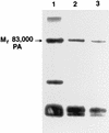
FIG. 1
FIG. 1
SDS-PAGE analysis of ΔSterne-1(pPA102)CR4 culture supernatants from complete, reduced-yeast-extract, and reduced-tryptone media. Lanes: 1, complete medium with 33 g of tryptone/liter and 20 g of yeast extract/liter; 2, complete reduced yeast extract medium with 33 g of tryptone/liter and 5 g of yeast extract/liter; 3, complete reduced tryptone medium with 8.25 g of tryptone/liter and 20 g of yeast extract/liter. A 1-ml sample was removed from each culture at late log to early stationary phase. The samples were sterile filtered, buffer exchanged, and concentrated to equivalent volumes. Equivalent volumes were loaded for SDS-PAGE analysis.
FIG. 2
FIG. 2
Physical-chemical parameters from aerobic ΔSterne-1(pPA102)CR4 fermentation. Symbols: open circles, DO2; solid line, pH; open squares, agitation; solid squares, percent oxygen. The tryptone addition during the fermentation is indicated by the arrow above the percent oxygen data at 200 min. The temperature range was 36.7 to 37.4°C. Pressure was constant at 2.0 lb/in2, and the sparge rate was 1 vol/vol/min. The sharp positive spike in the DO2 values at 350 min was due to the addition of antifoam KFO673.
FIG. 3
FIG. 3
PA production versus cell density. Symbols: solid circles, growth data 7 July fermentation; open circles, growth data 4 August fermentation; solid triangles, PA yield data 7 July fermentation; open triangles, PA yield data 4 August fermentation. Equivalent volumes were sampled from the 20-liter fermentor for each EFT. The OD600 was determined for each EFT, and the samples were sterile filtered. The filtrates were desalted and concentrated to equivalent final volumes before a 1:1 dilution with twofold-concentrated SDS solubilization buffer and analysis by SDS-PAGE. Coomassie blue-stained gels were digitally scanned as described in Materials and Methods, and amounts of Mr 83,000 PA were determined for each EFT.
FIG. 4
FIG. 4
SDS-PAGE results reflecting the time course of PA production from aerobic fermentation of ΔSterne-1(pPA102)CR4. Lanes: 1, molecular weight standards; 2, EFT 1 h; 3, EFT 2 h; 4, EFT 3 h; 5, EFT 4 h; 6, EFT 5 h; 7, EFT 6 h; 8, immunoblot of EFT 6 h developed with a pool of four monoclonal antibodies. Equivalent sample volumes were taken for each EFT. Samples were sterile filtered, and the filtrate was desalted, lyophilized, and resuspended in buffer to the same final volume before a 1:1 dilution with twofold-concentrated SDS solubilization buffer and analysis by SDS-PAGE. Equal volumes of each sample were applied to the gel. The immunoblot was developed with 4-chloro-1-naphthol after incubation with goat-anti mouse antibody linked to horseradish peroxidase. Bio-Rad low-molecular-weight standards with the following Mrs were used: phosphorylase b, 97,400; serum albumin, 66,200; ovalbumin, 45,000; carbonic anhydrase, 31,000; trypsin inhibitor, 21,500; and lysozyme, 14,400.
FIG. 5
FIG. 5
UV-spectrophotometric and SDS-PAGE analysis of the diafiltration permeate. Symbols: solid circles, A260; open circles, A280. Permeate from the Amicon 30-kDa-cutoff spiral-wound cartridge during the first diafiltration step was collected, diluted with the diafiltration buffer, and analyzed by UV spectroscopy at 280 and 260 nm. The spectrophotometer was blanked against the diafiltration buffer. Values at 350 and 320 nm were measured to confirm that sample turbidity was negligible. The inset shows a Coomassie blue-stained SDS-PAGE gel of a 10-fold concentrate of the diafiltration permeate collected at the outset of the diafiltration step.
FIG. 6
FIG. 6
SDS-PAGE analysis of PA purity after chromatographic purification steps. Lanes: 1, molecular weight standards; 2, PA after Macro-Prep 50 Q chromatography; 3, PA after HQ chromatography; 4, PA after QE chromatography; 5, PA after ether HIC chromatography. PA samples from each purification step were desalted, concentrated, and solubilized in SDS buffer. The total protein loaded in lanes 2 to 5 was 2 μg. Molecular weight standards were as in Fig. 4.
FIG. 7
FIG. 7
Determination of PA purity by SDS capillary electrophoresis. PA samples were analyzed after QE (A) or ether HIC (B) chromatography steps. The samples were desalted and concentrated to 3 to 4 mg/ml before being solubilized with SDS as described in Materials and Methods. The samples were applied by electrokinetic injection at −5 kV for 5 to 10 s. Separations were performed at −10 kV for 15 min, and the A215 was monitored.
FIG. 8
FIG. 8
Reversed-phase HPLC analysis of PA. A 100-μg sample of POROS QE-purified PA was subjected to reversed-phase HPLC analysis. The three main absorbance peaks labeled 1 were collected and pooled, and peak 2 was collected separately. Fractions collected for SDS-PAGE were immediately neutralized by adding 20 mM HEPES (pH 7.3) and diluting 1:1 with MQ water before concentrating. The results of Coomassie blue-stained SDS-PAGE are shown in the inset. The numbering of the inset lanes corresponds to the peak labeling. The Mr 80,000 contaminant in lane 1 is identified by an arrow to the left of the inset.
FIG. 9
FIG. 9
Immunoblot analysis of the total impurities in Mr 83,000 PA purified by QE anion-exchange chromatography. The pooled protein contaminants from three separate lots of PA were purified by reversed-phase HPLC, separated by SDS-PAGE, and transferred electrophoretically to nitrocellulose membranes. The immunoreactive bands were detected with the ECL reagent and a pool of monoclonal antibodies as described in Materials and Methods.

Similar articles

Cited by

References

    1. Arbigen M V, Bulthuis B A, Schultz J, Crabb D. Fermentation of Bacillus. In: Sonenshein A L, Hoch J A, Losick R, editors. Bacillus subtilis and other gram-positive bacteria. Washington, D.C: American Society for Microbiology; 1993. pp. 871–895.
    1. Bruecker R, Shoseyov O, Doi R H. Multiple active forms of a novel serine protease from Bacillus subtilis. Mol Gen Genet. 1990;221:486–490. - PubMed
    1. Dubois M, Gilles K A, Hamilton T K, Rebers P A, Smith F. Colorimetric method for determination of sugars and related substances. Anal Chem. 1956;28:350–353.
    1. Ezzell J W, Abshire T G. Serum protease cleavage of Bacillus anthracis protective antigen. J Gen Microbiol. 1992;138:543–549. - PubMed
    1. Farchaus W, Ribot W J, Downs M B, Ezzell J W. Purification and characterization of the major surface array protein from the avirulent Bacillus anthracis strain Delta Sterne-1. J Bacteriol. 1995;177:2481–2489. - PMC - PubMed

MeSH terms

LinkOut - more resources