. 1998 Apr 1;18(7):2321-34.
doi: 10.1523/JNEUROSCI.18-07-02321.1998.

Upregulation of L-type Ca2+ channels in reactive astrocytes after brain injury, hypomyelination, and ischemia

Affiliations

Upregulation of L-type Ca2+ channels in reactive astrocytes after brain injury, hypomyelination, and ischemia

R E Westenbroek et al. J Neurosci. .

Abstract

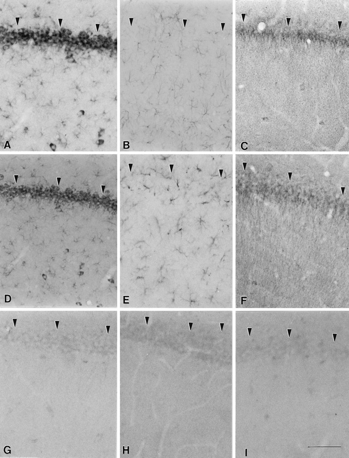
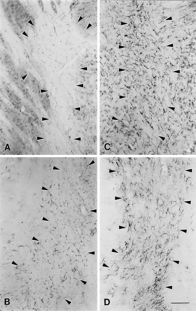
Anti-peptide antibodies that specifically recognize the alpha1 subunit of class A-D voltage-gated Ca2+ channels and a monoclonal antibody (MANC-1) to the alpha2 subunit of L-type Ca2+ channels were used to investigate the distribution of these Ca2+ channel subtypes in neurons and glia in models of brain injury, including kainic acid-induced epilepsy in the hippocampus, mechanical and thermal lesions in the forebrain, hypomyelination in white matter, and ischemia. Immunostaining of the alpha2 subunit of L-type Ca2+ channels by the MANC-1 antibody was increased in reactive astrocytes in each of these forms of brain injury. The alpha1C subunits of class C L-type Ca2+ channels were upregulated in reactive astrocytes located in the affected regions in each of these models of brain injury, although staining for the alpha1 subunits of class D L-type, class A P/Q-type, and class B N-type Ca2+ channels did not change from patterns normally observed in control animals. In all of these models of brain injury, there was no apparent redistribution or upregulation of the voltage-gated Ca2+ channels in neurons. The upregulation of L-type Ca2+ channels in reactive astrocytes may contribute to the maintenance of ionic homeostasis in injured brain regions, enhance the release of neurotrophic agents to promote neuronal survival and differentiation, and/or enhance signaling in astrocytic networks in response to injury.


Figures

Fig. 1.
Fig. 1.
Distribution of L-type Ca2+channels and GFAP in the CA3 region of the hippocampus of kainate acid-injected or in uninjected rats. A, Section stained with MANC-1 illustrating the distribution of the α2subunit of L-type Ca2+ channels in the cell soma, proximal dendrites, and astrocytic processes of a control rat.B, Expression of MANC-1 in reactive astrocytes and remaining neurons after kainate acid injection. C, Control section stained with anti-GFAP. D, Section stained with anti-GFAP illustrating the presence of reactive astrocytes after kainate acid injection. E, Section stained with anti-CNC1 illustrating the pattern of distribution of the α1 subunit of class C L-type Ca2+channels in control rats. F, Section from animal injected with kainate acid illustrating the presence of anti-CNC1 staining in reactive astrocytes and the remaining neurons. Scale bar, 200 μm.
Fig. 2.
Fig. 2.
Distribution of L-type Ca2+channels and GFAP in the CA1 region of the hippocampus of kainate acid-injected and uninjected rats. A, Section stained with MANC-1 illustrating the distribution of the α2subunit of L-type Ca2+ channels along neurons and astrocytic processes of a control rat. B, Section from a control animal stained with anti-GFAP. C, Section labeled with anti-CNC1 illustrating the pattern of distribution of the α1 subunit of class C L-type Ca2+channels in control rats. D, Section from kainate acid-injected animal stained with MANC-1 to illustrate that no changes occurred in the distribution of the α2 subunit in neurons or astrocytes. E, Section stained with GFAP demonstrating that the pattern of distribution of this protein is unaltered in the CA1 region after kainate acid injection.F, Section from the same experimental animal stained with anti-CNC1 illustrating that the distribution of the α1 subunit of L-type Ca2+ channels remains unaltered in the CA1 region. G, Control section demonstrating the lack of staining observed when MANC-1 is preabsorbed with purified Ca2+ channels. H, Control section in which the primary antiserum was replaced by normal mouse IgG. I, Control section in which the primary antibody was replaced by normal rabbit serum. Arrowheadspoint to the pyramidal cell body layer. Scale bar, 100 μm.
Fig. 3.
Fig. 3.
Distribution of class A, B, and D Ca2+ channels in the CA3 region of the hippocampus after kainic acid injection. A, Section stained with anti-CND1 antibodies illustrating the localization of class D channels in cell bodies of the remaining neurons and not in the pyramidal neurons affected by the kainic acid injection (arrows) or in reactive astrocytes. B, Section incubated with anti-CNA1 antibodies showing the lack of staining in the pyramidal neurons affected by the kainic acid injection, as compared with the normal pattern of staining along the length of the remaining pyramidal cells. C, Section stained with anti-CNB2 antibody illustrating the normal pattern of class B distribution in the remaining neurons after kainic acid injection. D, Control section in which the primary antibody was omitted. Scale bar, 200 μm.
Fig. 4.
Fig. 4.
Localization of MANC-1, GFAP, and anti-CNC1 in wild-type and shiverer mice. A, Section incubated with MANC-1 demonstrating the presence of L-type Ca2+ channels in the neuronal cell bodies and in the processes of astrocytes found in the corpus callosum of wild-type mice (between arrows). B, Section stained with MANC-1 illustrating the presence of L-type Ca2+channels in reactive astrocytes found in the corpus callosum ofshiverer mice. C, Tissue from a wild-type mouse stained with anti-GFAP showing the pattern of astrocyte staining.D, Tissue section from a shiverer mouse stained with anti-GFAP illustrating numerous reactive astrocytes in the corpus callosum. E, Higher magnification of anti-GFAP staining in wild-type mice. F, Higher magnification of anti-GFAP staining in shiverer mice illustrating the enlarged somas and large, thick processes of reactive astrocytes, as compared with wild-type mice. Scale bars: 200 μm inA–D; 100 μm in E, F.
Fig. 5.
Fig. 5.
Localization of MANC-1 and GFAP in the internal capsule of wild-type and shiverer mice.A, Tissue section from wild-type mouse stained with MANC-1 illustrating low levels of expression of L-type Ca2+ channels in astrocytes located in the internal capsule. B, Tissue section stained with anti-GFAP that shows the distribution L-type Ca2+ channels in wild-type mice. C, Micrograph illustrating the staining of MANC-1 in reactive astrocytes located in the internal capsule of ashiverer mouse. D, Section from ashiverer mouse illustrating the increased staining observed in the internal capsule with anti-GFAP antibodies.Arrowheads outline the region of the internal capsule in wild-type and mutant mice. Scale bar, 200 μm.
Fig. 6.
Fig. 6.
Distribution of L-type Ca2+channels and GFAP in the cerebellum of wild-type andshiverer mice. A, MANC-1 staining in a section from the cerebellum of a wild-type mouse. B, MANC-1 staining in a section from the cerebellum of ashiverer mouse illustrating staining in astrocytic processes in the corpus medullare region. C, Section from a wild-type mouse stained with anti-GFAP antibodies.D, Section stained with anti-GFAP antibodies illustrating the presence of reactive astrocytes in the white matter of the cerebellum of a shiverer mouse. E, Section stained with anti-CNC1 antibodies showing the pattern of class C Ca2+ channel staining in the corpus medullare of the cerebellum of a wild-type mouse. F, Section from ashiverer mouse demonstrating the staining of reactive astrocytes located in the corpus medullare of the cerebellum with anti-CNC1 antibodies. Arrowheads outline the region of the corpus medullare region in wild-type and mutant mice. Scale bar, 100 μm.
Fig. 7.
Fig. 7.
Expression of L-type Ca2+channels in reactive astrocytes after mechanical lesions.A, Photomicrographs showing abundant reactive astrocytes stained by MANC-1 in the striatum 3 weeks after a stab wound.B, Photomicrographs showing abundant reactive astrocytes stained by MANC-1 in the ventro-posterior nucleus of the thalamus 3 weeks after aspiration of the somatosensory cortex. C, Double immunofluorescent staining of MANC-1 (Texas Red) and GFAP (D, FITC) of reactive astrocytes in the superficial layers of the neocortex 3 weeks after thermal lesions of the adjacent neocortex. Scale bars: 200 μm in A; 50 μm inB.
Fig. 8.
Fig. 8.
L-type Ca2+ channels in tanycytes. Double immunofluorescent staining of MANC-1 (A, Texas Red) and GFAP (B, FITC) in tanycytes of the median eminence. Scale bar, 50 μm.
Fig. 9.
Fig. 9.
Expression of MANC-1 immunoreactivity in astrocytes in forebrain nuclei after ischemic injury. A, B, Double immunofluorescent staining of MANC-1 (A, Texas Red) and GFAP (B, FITC) in reactive astrocytes in the CA1 sector of the hippocampus 7 d after experimental ischemia, as described in Materials and Methods. Thearrows point to examples of reactive astrocytes double-stained for MANC-1 and GFAP. C, D, Reactive astrocytes expressing L-type Ca2+ channels stained by MANC-1 (C, Texas Red) and GFAP (D, FITC) in the dorsolateral sector of the striatum 7 d after ischemia. The arrows point to two representative examples. Scale bar, 30 μm.

Similar articles

Cited by

References

    1. Ahlijanian MK, Westenbroek RE, Catterall WA. Subunit structure and localization of dihydropyridine-sensitive Ca2+ channels in mammalian brain, spinal cord, and retina. Neuron. 1990;4:819–832. - PubMed
    1. Aicardi G, Schwartzkroin PA. Suppression of epileptiform burst discharges in CA1 neurons of rat hippocampal slices by the organic Ca2+ channel blocker, verapamil. Exp Brain Res. 1990;81:288–296. - PubMed
    1. Andersson H, Luthman J, Oslon L. Trimethyltin-induced expression of GABA and vimentin immunoreactivities in astrocytes of the rat brain. Glia. 1994;11:378–382. - PubMed
    1. Astrup J, Norberg K. K+ activity in cerebral cortex in rats during progressive severe hypoglycemia. Brain Res. 1976;103:418–423. - PubMed
    1. Ballarin M, Ernfors P, Linderfors N, Persson H. Hippocampal damage and kainic acid injection induce a rapid increase in mRNA for BDNF and NGF in the rat brain. Exp Neurol. 1991;114:35–43. - PubMed

Publication types

MeSH terms