. 1998 Apr 1;18(7):2412-22.
doi: 10.1523/JNEUROSCI.18-07-02412.1998.

Honeybee blue- and ultraviolet-sensitive opsins: cloning, heterologous expression in Drosophila, and physiological characterization

Affiliations

Honeybee blue- and ultraviolet-sensitive opsins: cloning, heterologous expression in Drosophila, and physiological characterization

S M Townson et al. J Neurosci. .

Abstract

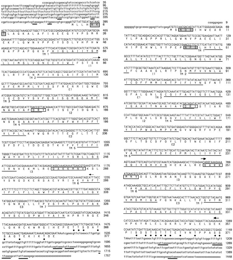
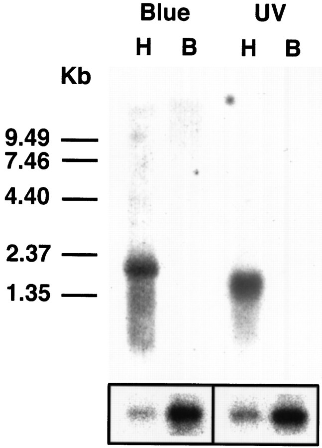
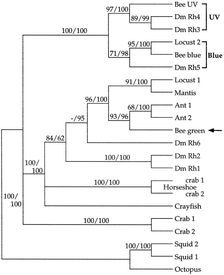
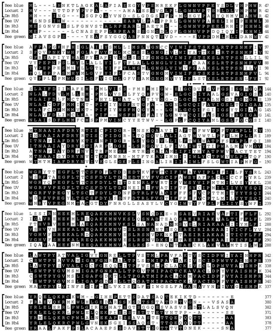
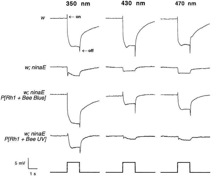
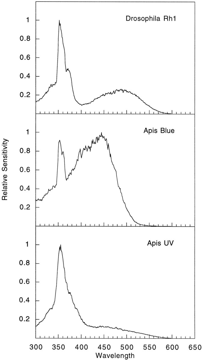
The honeybee (Apis mellifera) visual system contains three classes of retinal photoreceptor cells that are maximally sensitive to light at 440 nm (blue), 350 nm (ultraviolet), and 540 nm (green). We performed a PCR-based screen to identify the genes encoding the Apis blue- and ultraviolet (UV)-sensitive opsins. We obtained cDNAs that encode proteins having a high degree of sequence and structural similarity to other invertebrate and vertebrate visual pigments. The Apis blue opsin cDNA encodes a protein of 377 amino acids that is most closely related to other invertebrate visual pigments that are thought to be blue-sensitive. The UV opsin cDNA encodes a protein of 371 amino acids that is most closely related to the UV-sensitive Drosophila Rh3 and Rh4 opsins. To test whether these novel Apis opsin genes encode functional visual pigments and to determine their spectral properties, we expressed them in the R1-6 photoreceptor cells of blind ninaE mutant Drosophila, which lack the major opsin of the fly compound eye. We found that the expression of either the Apis blue- or UV-sensitive opsin in transgenic flies rescued the visual defect of ninaE mutants, indicating that both genes encode functional visual pigments. Spectral sensitivity measurements of these flies demonstrated that the blue and UV visual pigments are maximally sensitive to light at 439 and 353 nm, respectively. These maxima are in excellent agreement with those determined previously by single-cell recordings from Apis photoreceptor cells and provide definitive evidence that the genes described here encode visual pigments having blue and UV sensitivity.


Figures

Fig. 1.
Fig. 1.
Nucleotide sequences and deduced amino acid sequences of the Apis blue-sensitive (left) and UV-sensitive (right) opsins. Nucleotides are numbered in the 5′- to 3′-direction. The deduced amino acid sequences are shown below the nucleotide sequences in single letter code. A single major open reading frame of 1131 bp is present in the blue opsin cDNA encoding a protein of 377 amino acids. The UV opsin major open reading frame is 1113 bp long and encodes a protein of 371 amino acids. Seven potential transmembrane (TM) domains areunderlined (Kyte and Doolittle, 1982). The stop codons in the 5′-untranslated region and the putative polyadenylation signals upstream of the polymeric dA tract in the 3′-untranslated region are also underlined (thick lines). The blue opsin cDNA contains four putative polyadenylation signals beginning at nucleotides 1627, 1634, 1714, and 1726. The AUUAAA polyadenylation signal is the most common variant of the AAUAAA polyadenylation signal (Swimmer and Shenk, 1985). The 3′-untranslated region of the UV cDNA has polyadenylation signals beginning at nucleotides 1231, 1264, and 1417. There are three additional out-of-frame stop codons in the 5′-untranslated region of the blue opsin cDNA. Potential sequences for G-protein binding sites, DRY and QAKKMNV, as mentioned in the Results, are indicated by open boxes. Potential glycosylation sites in the N terminals are indicated by shaded boxes. Possible Ser and Thr phosphorylation sites are indicated bysolid circles. The intracellular (I) and extracellular (E) loops are indicated below the amino acid sequence. The original 606 bp blue opsin fragment identified by PCR from nucleotides 627 to 1232 is indicated between thearrows. The 310 bp fragment identified by PCR from nucleotide 777 to 1086 for the UV gene is indicated similarly.
Fig. 2.
Fig. 2.
Expression of the Apis blue- and UV-sensitive opsin genes. Northern analysis using theApis blue 1–1A and UV 7–1 cDNAs as probes revealed that the genes are transcribed as 2.2 and 1.7 kb mRNAs, respectively. These transcripts were present in the heads (H) but not in the bodies (B) of adult bees. The lower boxshows the same filter probed with a 460 bp fragment of the honeybee cytochrome C oxidase subunit 1 gene and demonstrates that mRNA is present in the samples prepared from both bee heads and bodies. The size of the polycistronic transcript that contains the control probe sequence is 1.9 kb. Size markers in kb are shown on theleft.
Fig. 3.
Fig. 3.
Phylogenetic relationships between theApis blue- and UV-sensitive opsins and other known visual pigments. The Apis blue- and UV-sensitive opsins fall into two different groups that are thought to be sensitive to blue and UV light, respectively. The blue-sensitive group includes the recently identified Locust 2 and Drosophila Rh5, whereas the UV-sensitive group includes the known UV-sensitive Drosophila Rh3 and Rh4pigments. The relative position of the putative Apisgreen-sensitive opsin is indicated with an arrow. This tree highlights that, although these pigments have been identified in highly divergent species, they are nonetheless most closely related to visual pigments from other organisms believed to have similar spectral properties. Amino acid sequences were aligned using the program ClustalW (Thompson et al., 1994). The regions of the alignment corresponding to amino acids 27–361 of the bee blue and amino acids 18–353 of the bee UV opsin were used for the analysis. Thirty-four residues of the alignment at the N terminal and 141 residues at the C terminal (including the Pro-rich repeats in the cephalopod opsins) were excluded from the analysis because of possible alignment ambiguities arising from substantial differences in sequence length (although including the terminals in the phylogenetic analysis did not significantly alter tree topology). Two types of phylogenetic analysis were used: maximum parsimony (Swofford, 1991) and neighbor-joining (Saitou and Nei, 1987). Both were performed using PAUP* 4.0 running on a Power personal computer (test versions kindly provided by D. L. Swofford). Robustness of the results was assessed using bootstrap analysis (Felsenstein, 1985). One hundred bootstrap replications, with five random additions each, were done using unweighted parsimony. One hundred bootstrap replications were done using neighbor-joining, with tree-bisection reconnection branch swapping to ensure finding the shortest tree. The results of these two analyses were in agreement, although levels of support for particular nodes differed. The tree was rooted using vertebrate opsin sequences (data not shown). Nodes with bootstrap values below 65 for both analyses were collapsed. Bootstrap values are shown above each node (parsimony/neighbor-joining). Abbreviations and GenBank accession numbers for the sequences used in the construction of the tree are as follows: Apis mellifera (Bee blue, AF004168; Bee UV, AF004169; Bee green, U26026); Camponotus abdominalis(Ant 1, U32502); Cataglyphis bombycina(Ant 2, U32501); Drosophila melanogaster(Dm Rh1, P06002; Dm Rh2, P08099;Dm Rh3, P04950; Dm Rh4, P29404; Dm Rh5, U67905; Dm Rh6, Z86118); Hemigrapsus sanguineus (Crab 1, D50583; Crab 2, D50584); Limulus polyphemus (lateral eye opsin Horse-shoe crab 1, L03781; ocellar opsinHorseshoe crab 2, L03782); Loligo forbesi (Squid 1, X56788); Octopus dofleini(Octopus, X07797); Procambarus clarkii(Crayfish, S53494); Schistocerca gregaria(Locust 1, X80071; Locust 2, X80072);Sphodromantis sps (Mantis, X71665); andTodarodes pacificus (Squid 2,X70498).
Fig. 4.
Fig. 4.
Amino acid sequence alignment between theApis blue- and UV-sensitive opsins and other related pigments. The sequences are grouped as indicated in Figure 3. The pigments thought to be blue-sensitive include Drosophila Rh5 and Locust 2, whereas the UV-sensitive opsins include Drosophila Rh3 and Rh4. The putative Apis green opsin is indicated on thelower line for comparison. Consensus amino acids areblackened. The potential transmembrane segments are indicated with brackets over thesequences. Highly conserved amino acids are indicated with an asterisk and are discussed in the Results. These include the Lys in TM 7 and a pair of Cys at the beginning of TM 3 and between TM 4 and TM 5. The residue corresponding to the vertebrate counterion (Tyr in the blue and green pigments vs Phe in the UV pigments) in TM 3 is also indicated.
Fig. 5.
Fig. 5.
Electroretinogram recordings of transgenic flies expressing the Apis blue- and UV-sensitive opsins. Eachcolumn shows the light response to a 1 sec flash at different wavelengths of light, 350 nm (left), 430 nm (middle), and 470 nm (right). Eachrow shows the ERG recording from a different genetic background. w1118 flies (top) respond to light at all three wavelengths with a robust depolarization and on and off transients (see Results).w1118; ninaE17 flies (second from the top), which lack the ninaE (Rh1) opsin of the R1–6 photoreceptor cells, lack the on and off transients and have a severely reduced receptor potential at all three wavelengths. Transgenic flies expressing the Apis blue opsin (third from the top;w1118; ninaE17 P[Rh1 + Bee Blue]) show a robust response to light at all wavelengths, with a complete recovery of the depolarization and transients. Transgenic flies expressing the Apis UV opsin (fourth fromtop; w1118; ninaE17 P[Rh1 + Bee UV]) show a normal depolarization and transients in response to UV stimulation. The amplitudes of the ERG response are not comparable between different strains because of differences in expression levels of the transgenes, nor are they comparable at different wavelengths because of differences in stimulus intensity. For most recordings, light intensity was attenuated 3 OD, resulting in intensities of ∼0.22, 1.3, and 2.7 μW/cm2 at 350, 430, and 470 nm, respectively. With the exception of the response fromw flies (which was recorded at 3 OD as indicated above), all of the responses at 350 nm were recorded with a light intensity attenuated 1 OD, corresponding to a light intensity of 21 μW/cm2.
Fig. 6.
Fig. 6.
Spectral sensitivity recordings of flies expressing the Apis blue- and UV-sensitive opsins.Top, The spectral sensitivity profile of a white-eyed fly (w1118) that expressesDrosophila Rh1 in the R1–6 photoreceptor cells. These animals display a dual peak of sensitivity. The peak in the UV is attributable to the effect of a sensitizing pigment that absorbs in the UV and transfers the energy of the photon to the Rh1 rhodopsin to activate it. There is also a prominent peak of sensitivity in the blue region with a maximum at 479 nm. Middle, The spectral sensitivity profile of flies expressing the Apisblue-sensitive opsin in a genetic background in which the endogenous opsin expressed in the R1–6 photoreceptor cells has been deleted (w1118; ninaE17 P[Rh1 + Bee Blue]). Much like the white-eyed control animals, flies expressing theApis blue-sensitive opsin have a dual peak of sensitivity, which we believe results from the coupling of theApis blue opsin to the UV-sensitizing pigment in a manner similar to that of Rh1. The principal peak of sensitivity is in the blue region with a maximum at 439 nm. Lower, The spectral sensitivity profile of flies expressing theApis UV-sensitive opsin (w1118; ninaE17 P[Rh1 + Bee UV]). These flies have a single peak of sensitivity in the UV region with a maximum at 353 nm. The spectral sensitivity of the ninaEhost strain is not detectable by these methods, because the response levels (as shown in Fig. 5) are not large enough to meet the criterion of the recording paradigm. These results are in excellent agreement with published intracellular recordings of the Apisblue- and UV-sensitive photoreceptors (Menzel et al., 1986) and provide conclusive evidence that the cloned genes encode biologically active visual pigments having the indicated spectral properties.

Similar articles

Cited by

References

    1. Asenjo AB, Rim J, Oprian DD. Molecular determinants of human red/green color discrimination. Neuron. 1994;12:1131–1138. - PubMed
    1. Baldwin JM. The probable arrangement of the helices in G protein-coupled receptors. EMBO J. 1993;12:1693–1703. - PMC - PubMed
    1. Bellingham J, Wilkie SE, Morris AG, Bowmaker JK, Hunt DM. Characterisation of the ultraviolet-sensitive opsin gene in the honey bee, Apis mellifera. Eur J Biochem. 1997;243:775–781. - PubMed
    1. Bentrop J, Schwab K, Pak WL, Paulsen R. Site-directed mutagenesis of highly conserved amino acids in the first cytoplasmic loop of Drosophila Rh1 opsin blocks rhodopsin synthesis in the nascent state. EMBO J. 1997;16:1600–1609. - PMC - PubMed
    1. Bernard GD, Remington CL. Color vision in Lycaena butterflies: spectral tuning of receptor arrays in relation to behavioral ecology. Proc Natl Acad Sci USA. 1991;88:2783–2787. - PMC - PubMed

Publication types

Associated data

LinkOut - more resources