. 1998 Apr 1;18(7):2423-36.
doi: 10.1523/JNEUROSCI.18-07-02423.1998.

Role of actin in anchoring postsynaptic receptors in cultured hippocampal neurons: differential attachment of NMDA versus AMPA receptors

Affiliations

Role of actin in anchoring postsynaptic receptors in cultured hippocampal neurons: differential attachment of NMDA versus AMPA receptors

D W Allison et al. J Neurosci. .

Abstract

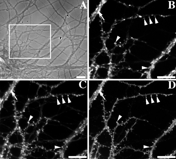
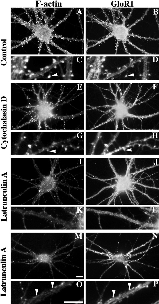
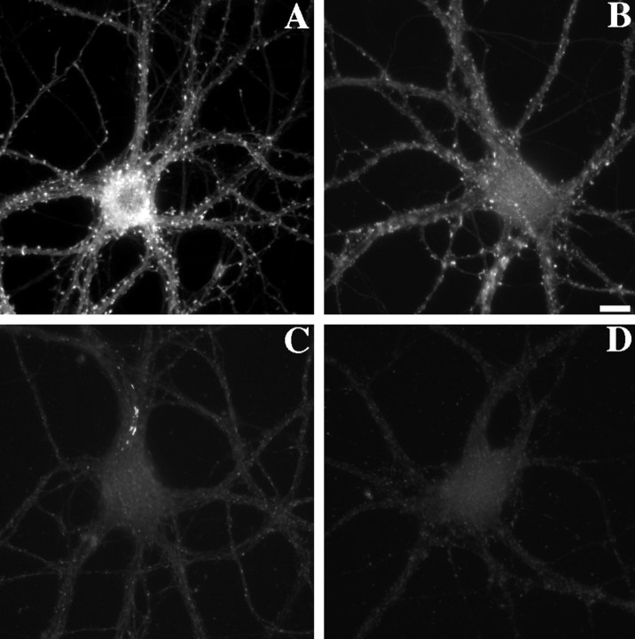
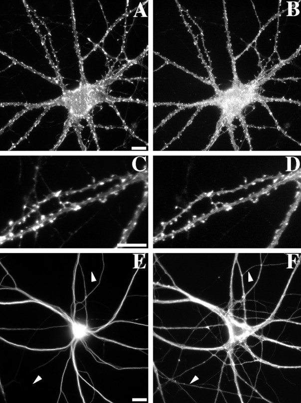
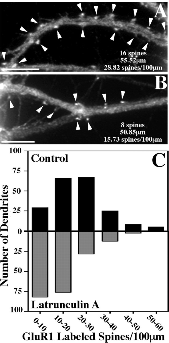
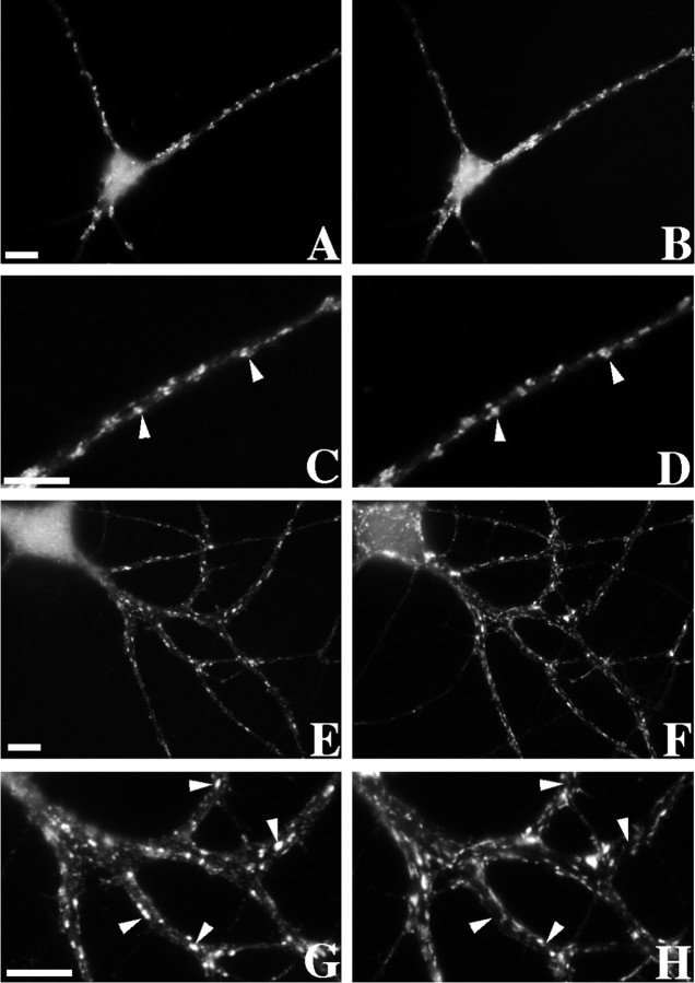
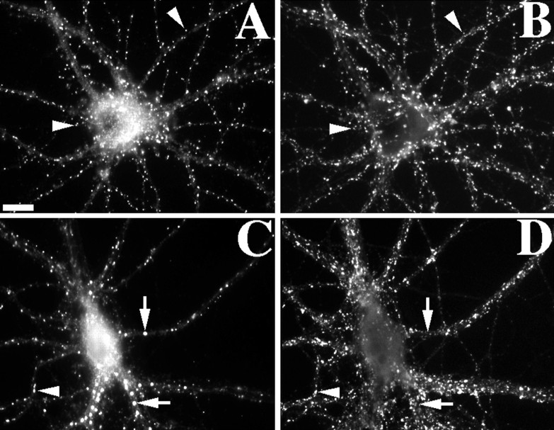
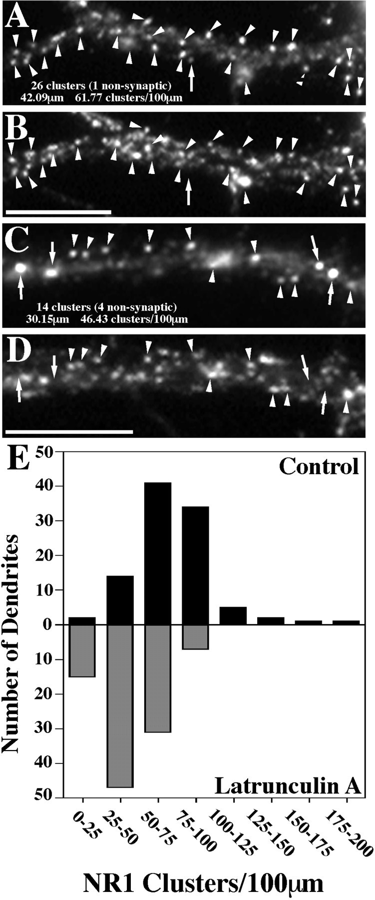
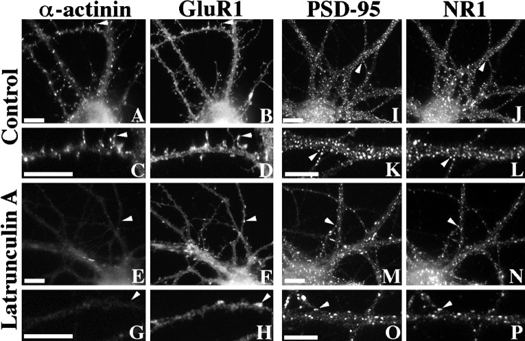
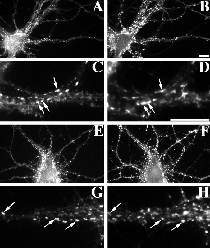
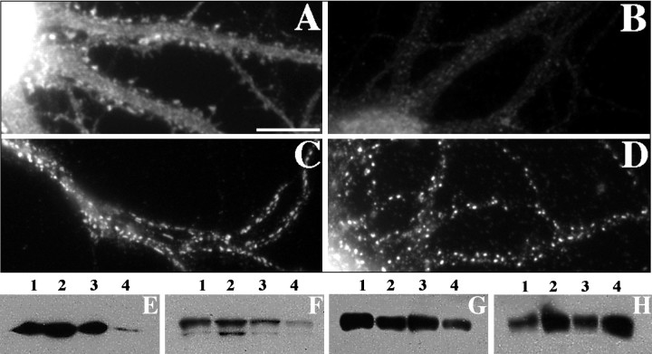
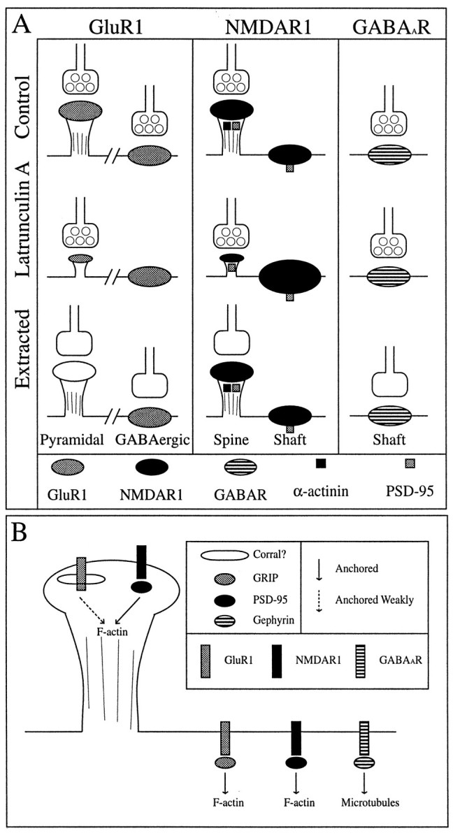
We used actin-perturbing agents and detergent extraction of primary hippocampal cultures to test directly the role of the actin cytoskeleton in localizing GABAA receptors, AMPA- and NMDA-type glutamate receptors, and potential anchoring proteins at postsynaptic sites. Excitatory postsynaptic sites on dendritic spines contained a high concentration of F-actin that was resistant to cytochalasin D but could be depolymerized using the novel compound latrunculin A. Depolymerization of F-actin led to a 40% decrease in both the number of synaptic NMDA receptor (NMDAR1) clusters and the number of AMPA receptor (GluR1)-labeled spines. The nonsynaptic NMDA receptors appeared to remain clustered and to coalesce in cell bodies. alpha-Actinin-2, which binds both actin and NMDA receptors, dissociated from the receptor clusters, but PSD-95 remained associated with both the synaptic and nonsynaptic receptor clusters, consistent with a proposed cross-linking function. AMPA receptors behaved differently; on GABAergic neurons, the clusters redistributed to nonsynaptic sites, whereas on pyramidal neurons, many of the clusters appeared to disperse. Furthermore, in control neurons, AMPA receptors were detergent extractable from pyramidal cell spines, whereas AMPA receptors on GABAergic neurons and NMDA receptors were unextractable. GABAA receptors were not dependent on F-actin for the maintenance or synaptic localization of clusters. These results indicate fundamental differences in the mechanisms of receptor anchoring at postsynaptic sites, both regarding the anchoring of a single receptor (the AMPA receptor) in pyramidal cells versus GABAergic interneurons and regarding the anchoring of different receptors (AMPA vs NMDA receptors) at a single class of postsynaptic sites on pyramidal cell dendritic spines.


Figures

Fig. 1.
Fig. 1.
Coclustering of GluR1 and F-actin at synapses on dendritic spines of cultured rat hippocampal neurons. A, Phase contrast image of a typical pyramidal neuron at 3 weeks in culture (boxed area shows region enlarged inB–D). B–D, Staining with an antibody against the synaptic vesicle protein SV2 to label presynaptic terminals (B), with rhodamine phalloidin to label F-actin (C), and with an antibody against the GluR1 subunit of the AMPA-type glutamate receptor (D). GluR1 and F-actin were present at high concentrations at some spiny synapses (arrowheads). In contrast, synapses on dendrite shafts of pyramidal neurons did not exhibit high concentrations of F-actin or of GluR1 (arrow); these may correspond to glutamatergic synapses lacking concentration of the AMPA receptor or to GABAergic synapses. Scale bars, 10 μm.
Fig. 2.
Fig. 2.
Disruption of neuronal F-actin and its effect on GluR1-labeled spines. Neurons were stained at 3 weeks in culture with rhodamine phalloidin to label F-actin (A,C, E, G, I,K, M, O) and with an antibody against GluR1 (B, D,F, H, J, L,N, P). The smaller boxes(C, D, G,H, K, L, O,P) show enlarged regions from the neurons above (arrowheads represent spines). There were many spines with concentrations of both F-actin and GluR1 in control neurons (A–D) and after treatment with 10 μg/ml cytochalasin D for 24 hr (E–H). Although much of the cortical actin was disrupted by cytochalasin D, the spines were still positive for both F-actin (E,G) and GluR1 (F,H). In contrast, after a 24 hr treatment with 5 μm latrunculin A, most of the F-actin was depolymerized (I, K) with a corresponding loss of GluR1-labeled spines (J, L). Some neurons exhibited apparently “deflated” spines after latrunculin A treatment, protrusions close to the shafts lacking F-actin (M, O) but still containing concentrations of GluR1 (N, P). Scale bars, 10 μm.
Fig. 3.
Fig. 3.
Time course of the effects of latrunculin A in disrupting actin polymers. Hippocampal neurons were treated at 3 weeks in culture with 5 μm latrunculin A, fixed at time 0 (A) or after 2 hr (B), 9 hr (C), or 24 hr (D) of treatment, and stained for F-actin with rhodamine phalloidin. After 2 hr of latrunculin A treatment, cortical F-actin was reduced but still detectable and in particular was concentrated in dendritic spines. After 9 hr of latrunculin A treatment, most of the F-actin was depolymerized, although there were still some dendrite shaft regions (C) and a few spines containing F-actin (data not shown). After latrunculin A treatment for 24 hr, the neurons were almost devoid of F-actin staining, and F-actin-labeled spines were not observed. The images in A–D were taken at the same exposure and scaled equally to preserve the differences in F-actin staining. Scale bar, 10 μm.
Fig. 4.
Fig. 4.
Reversal of latrunculin A effects and maintenance of neuronal polarity. Hippocampal neurons were treated for 24 hr with latrunculin A (E, F) and in some cases were allowed to recover for an additional 24 hr in latrunculin A-free media (A–D; C andD show enlarged regions of A andB). A 24 hr reversal lead to a complete recovery of the normal F-actin staining pattern and GluR1 clustering on dendritic spines (F-actin, A, C; GluR1,B, D). As a control for the specificity of latrunculin A, the distributions of the microtubule-associated proteins MAP2 (a dendritic marker, E) and tau (an axonal marker, F) were shown to be unaffected by the 24 hr latrunculin A treatment. There was no difference in the staining patterns between latrunculin A-treated (E,F) and paired control (data not shown) neurons.Arrowheads indicate axons that are tau-positive and MAP2-negative. Scale bars, 10 μm.
Fig. 5.
Fig. 5.
GluR1-labeled spines decrease in number after latrunculin A treatment. The number of GluR1-labeled dendritic spines per 100 μm of dendrite length was counted for 200 control and 200 latrunculin A-treated dendrites. A, B, Typical counts for regions of control and latrunculin A-treated dendrites, respectively. Arrowheads represent clusters of GluR1 on dendritic spines. Many of the spines remaining after latrunculin A treatment resemble the smaller deflated spines that are typical of this treatment. C, These data were then compiled into a graph in histogram form, with the black bars representing control neurons and the gray bars the latrunculin A-treated neurons. Control neurons exhibited 21.68 ± 0.81 (mean ± SEM) spines per 100 μm, whereas latrunculin A-treated neurons had only 13.57 ± 0.64 (mean ± SEM) spines per 100 μm. This represents a significant decrease in spine number (t test, p< 0.0001). Scale bars, 10 μm.
Fig. 6.
Fig. 6.
GABAergic neurons exhibit nonsynaptic GluR1 clusters after actin depolymerization. A–D, A typical GABAergic neuron immunostained for GluR1 (A, enlarged region in C) and the synaptic vesicle protein SV2 (B, enlarged region in D) is shown. GluR1 formed clusters on the dendrite shafts opposite SV2-labeled terminals in control neurons (arrowheads in C,D). E–H, After latrunculin A treatment to depolymerize F-actin, GluR1 still formed clusters on GABAergic neurons (E, G). However, the GluR1 clusters were no longer localized to synaptic sites defined by SV2-labeled terminals (F, H) but appeared to be randomly distributed in dendrites at nonsynaptic sites (arrowheads in G,H). Scale bars, 10 μm.
Fig. 7.
Fig. 7.
NR1 clusters depend partially on F-actin for their synaptic localization. Control neurons (A,B) or latrunculin A-treated neurons (C,D) were immunolabeled for the essential NMDA receptor subunit NR1 (A, C) and for the synaptic marker synaptophysin (B, D). In control neurons, NR1 formed primarily synaptic clusters (94% synaptic) on both dendrite shafts and on spines (arrowheads). After treatment with latrunculin A, synaptic NR1 clusters were still present but reduced in number. There was also an apparent increase in the number of large nonsynaptic clusters located in dendrite shafts and cell bodies (arrows). All of these neurons were pretreated with APV from 14–21 d in culture to induce the synaptic NR1 pattern (see Rao and Craig, 1997). Scale bar, 10 μm.
Fig. 8.
Fig. 8.
NR1 clusters decrease in number after latrunculin A treatment. The number of NR1 clusters per 100 μm of dendrite length was obtained by quantifying data from 100 control and 100 latrunculin A-treated dendrites. A, B, Regions of a control dendrite stained for NR1 and synaptophysin, respectively.C, D, Regions of a latrunculin A-treated dendrite stained for NR1 and synaptophysin, respectively.Arrowheads represent clusters of receptor on both spines and the shaft of the dendrites that colocalize with synaptophysin and are therefore synaptic. Arrows show NR1 clusters that do not exhibit synaptophysin staining and are thus classified as nonsynaptic. E, The data were then compiled into a graph in histogram form, with theblack bars representing control neurons and thegray bars the latrunculin A-treated neurons. Control neurons exhibited 73.82 ± 2.72 (mean ± SEM) spines per 100 μm, whereas latrunculin A-treated neurons had only 44.60 ± 1.78 (mean ± SEM) spines per 100 μm. This represents a significant decrease in total cluster number (t test,p < 0.0001). The number of nonsynaptic clusters on the dendrites did not change after latrunculin A treatment (5.86 ± 0.38 to 5.96 ± 0.45), and so the total change represents a selective decrease in synaptic NR1 clusters. The number of nonsynaptic clusters also appeared to increase in the cell bodies, which were not included in the quantitation. Scale bars, 10 μm.
Fig. 9.
Fig. 9.
Differential effect of actin depolymerization on the NMDA receptor-interacting proteins α-actinin-2 and PSD-95. Control (A–D) or latrunculin A-treated (E–H) neurons were immunolabeled for both α-actinin-2 (A, C, E,G) and GluR1 (B, D,F, H). α-Actinin-2 was often concentrated in dendritic spines of control neurons, partially colocalizing with GluR1 (arrowheads inA–D) (Rao et al., 1998). After latrunculin A treatment, α-actinin-2 immunoreactivity was no longer clustered or associated with any remaining GluR1 clusters (arrowheads inE–H). In contrast, PSD-95 (I,K, M, O) colocalized closely with NR1 (J, L, N,P) in both control (I–L) and latrunculin A-treated (M–P) neurons (arrowheads). Scale bars, 10 μm.
Fig. 10.
Fig. 10.
GABAA receptors do not depend on F-actin for clustering or synaptic localization. Control neurons (A–D) or latrunculin A-treated neurons (E–H) were immunolabeled for the GABAA receptor β2/3 subunits (A,C, E, G) and the synaptic marker SV2 (B, D, F,H). The GABAA receptor distribution appeared to be unaffected by actin depolymerization. Typical elongated GABAA receptor clusters were present on shafts of both control and latrunculin A-treated neurons, and these were opposite synaptic terminals (arrows). Scale bars, 10 μm.
Fig. 11.
Fig. 11.
Detergent extraction to assess cytoskeletal anchoring of glutamate receptors. A, B, GluR1 staining of pyramidal neurons from the same culture taken at the same exposure either unextracted (A) or after extraction with Triton X-100 (B) is shown. Detergent extraction induced an obvious decrease in the amount of GluR1 immunoreactivity and a change in the distribution pattern including a complete loss of GluR1 immunoreactivity from dendritic spines.C, In contrast, detergent-extracted GABAergic neurons retained GluR1 clusters with a typical distribution pattern; these clusters were presumably at synaptic sites, although we were unable to confirm this directly because the synaptic markers SV2 and synaptophysin were extracted by Triton X-100 (data not shown).D, After Triton X-100 extraction, NR1 immunoreactivity on pyramidal neurons was also indistinguishable from that of unextracted neurons. Scale bar: A–D, 10 μm.E–H, Western blot analyses of unextracted control neurons (lane 1), control neurons extracted with 1% Triton X-100 (lane 2), latrunculin A-treated unextracted neurons (lane 3), and latrunculin A-treated, Triton X-100-extracted neurons (lane 4) are shown. Protein loading was normalized to cell number such that lanes 2 and 4 contain protein derived from twice as many neurons as lanes 1 and 3. These blots were probed with antibodies against actin (E), α-actinin-2 (F), GluR1 (G), and NR1 (H). Very little actin remained in the latrunculin A-treated, extracted neurons (E,lane 4), indicating that most of the F-actin was depolymerized by latrunculin A and therefore extractable. α-Actinin-2 was also nearly completely extractable after latrunculin A treatment (F); although it can also bind NMDA receptors, it apparently is highly dependent on F-actin for cytoskeletal attachment. Surprisingly, GluR1 was partially (∼75%) extractable with or without F-actin present (evident by the decreased signal in lanes 2 and 4 ofG relative to lanes 1 and3, despite the twofold greater loading of lanes 2 and 4). This result is consistent with the loss of GluR1 immunoreactivity from pyramidal neurons after extraction (B). NR1 (H), on the other hand, did not seem to be detergent extractable even with latrunculin A treatment (because the relative signal intensities correspond to the loading differences).
Fig. 12.
Fig. 12.
Diagrammatic summary of results (A) and model (B).A, In control neurons, GluR1 exhibits a cell type-specific synaptic distribution, spiny on pyramidal neurons and clustering on the shafts of GABAergic neurons. NMDAR1 shows both spiny and shaft clusters on the pyramidal neurons; in this case, some of the shaft clusters are nonsynaptic (represented by the lack of a presynaptic terminal). The spiny NR1 clusters are immunopositive for both PSD-95 and α-actinin-2, but the shaft NR1 clusters contain only PSD-95. GABAA receptors are found as synaptic shaft clusters on pyramidal neurons. After latrunculin A treatment to depolymerize actin, GluR1-labeled spines are decreased in number with the remaining spines being much smaller and devoid of F-actin. GluR1 shaft clusters on GABAergic neurons are no longer synaptic. Synaptic NR1 clusters are also decreased in number after actin depolymerization, and there appear to be more and/or larger nonsynaptic NR1 clusters. α-Actinin-2 becomes completely diffuse with the loss of F-actin, but PSD-95 remains coclustered with both synaptic and nonsynaptic NMDA receptors. The inhibitory GABAA receptor (GABAAR) is apparently unaffected by latrunculin A treatment. Detergent extraction leads to a complete loss of GluR1 on the spines of pyramidal neurons but has no affect on GluR1 clusters on the shafts of GABAergic neurons. Both NR1 (and its interacting proteins α-actinin-2 and PSD-95) and GABAA receptors are not readily detergent extractable but remain tightly anchored at presumptive synaptic sites (synaptophysin is readily extractable, but the receptor staining patterns remain unchanged).B, The above data lead to a model for distinct mechanisms for anchoring of neurotransmitter receptors to the cytoskeleton, not only between different receptor types at a single site but also for the same receptor within different cell types. Particularly for GluR1 clusters on spines, the mechanism is not well understood. Many possibilities exist: a weak, detergent-extractable interaction, a spectrin-based corral, preferential membrane addition, or some other mechanism. The same receptor is anchored in a different manner in GABAergic neurons, possibly by the PDZ protein GRIP. The NMDA receptor forms clusters on spines and dendritic shafts in the presence of PSD-95 whether α-actinin-2 is present or not, suggesting a more central role for PSD-95 in anchoring NMDA receptors and a possible modulatory role for α-actinin-2. The GABAAR, which colocalizes with gephyrin, may be anchored to the microtubule cytoskeleton through gephyrin.

Similar articles

Cited by

References

    1. Aoki C, Venkatesan C, Go CG, Mong JA, Dawson TM. Cellular and subcellular localization of NMDA-R1 subunit immunoreactivity in the visual cortex of adult and neonatal rats. J Neurosci. 1994;14:5202–5222. - PMC - PubMed
    1. Banker GA, Cowan WM. Rat hippocampal neurons in dispersed cell culture. Brain Res. 1977;126:397–425. - PubMed
    1. Bartlett WP, Banker GA. An electron microscopic study of the development of axons and dendrites by hippocampal neurons in culture. II. Synaptic relationships. J Neurosci. 1984;4:1954–1965. - PMC - PubMed
    1. Baude A, Nusser Z, Roberts JD, Mulvihill E, McIlhinney RA, Somogyi P. The metabotropic glutamate receptor (mGluR1 alpha) is concentrated at perisynaptic membrane of neuronal subpopulations as detected by immunogold reaction. Neuron. 1993;11:771–787. - PubMed
    1. Beck KA, Nelson WJ. The spectrin-based membrane skeleton as a membrane protein-sorting machine. Am J Physiol. 1996;270:C1263–C1270. - PubMed

Publication types

MeSH terms

LinkOut - more resources