. 1998 Apr 1;18(7):2506-19.
doi: 10.1523/JNEUROSCI.18-07-02506.1998.

Aquaporin-4 water channel protein in the rat retina and optic nerve: polarized expression in Müller cells and fibrous astrocytes

Affiliations

Aquaporin-4 water channel protein in the rat retina and optic nerve: polarized expression in Müller cells and fibrous astrocytes

E A Nagelhus et al. J Neurosci. .

Abstract

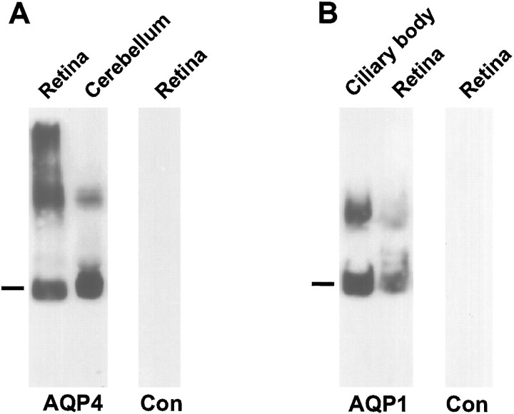
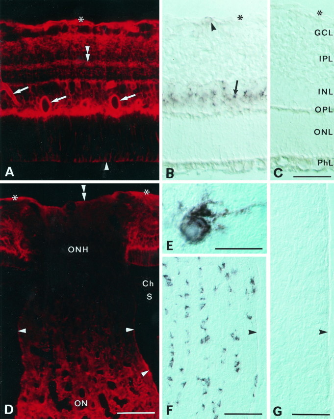
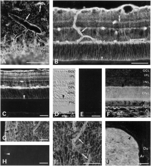
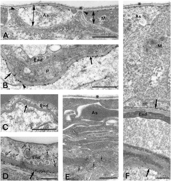
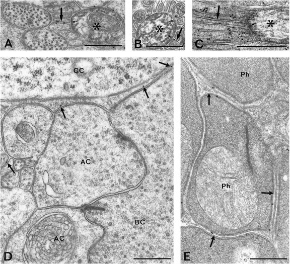
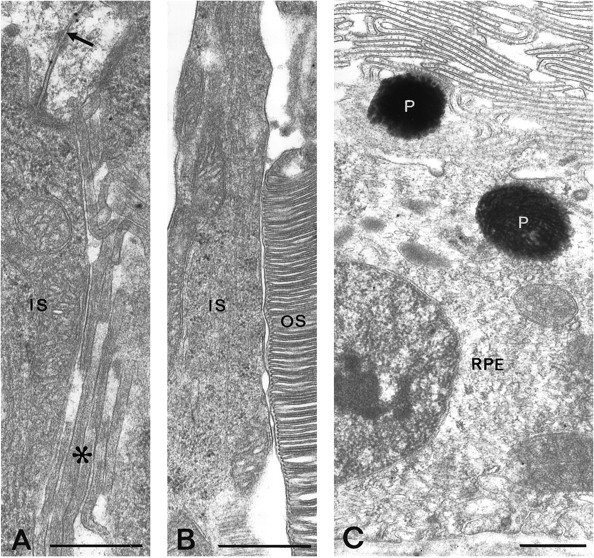
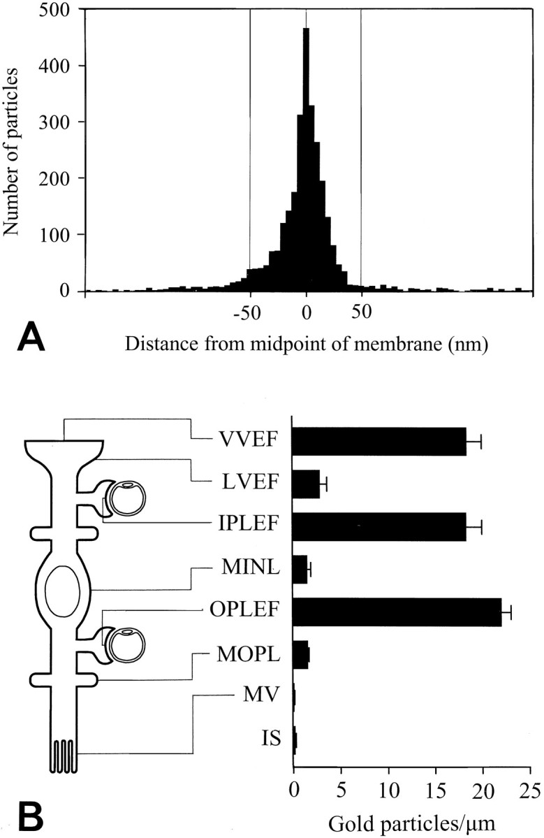
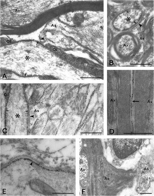
The water permeability of cell membranes differs by orders of magnitude, and most of this variability reflects the differential expression of aquaporin water channels. We have recently found that the CNS contains a member of the aquaporin family, aquaporin-4 (AQP4). As a prerequisite for understanding the cellular handling of water during neuronal activity, we have investigated the cellular and subcellular expression of AQP4 in the retina and optic nerve where activity-dependent ion fluxes have been studied in detail. In situ hybridization with digoxigenin-labeled riboprobes and immunogold labeling by a sensitive postembedding procedure demonstrated that AQP4 and AQP4 mRNA were restricted to glial cells, including MHller cells in the retina and fibrous astrocytes in the optic nerve. A quantitative immunogold analysis of the MHller cells showed that these cells exhibited three distinct membrane compartments with regard to AQP4 expression. End feet membranes (facing the vitreous body or blood vessels) were 10-15 times more intensely labeled than non-end feet membranes, whereas microvilli were devoid of AQP4. These data suggest that MHller cells play a prominent role in the water handling in the retina and that they direct osmotically driven water flux to the vitreous body and vessels rather than to the subretinal space. Fibrous astrocytes in the optic nerve similarly displayed a differential compartmentation of AQP4. The highest expression of AQP4 occurred in end feet membranes, whereas the membrane domain facing the nodal axolemma was associated with a lower level of immunoreactivity than the rest of the membrane. This arrangement may allow transcellular water redistribution to occur without inducing inappropriate volume changes in the perinodal extracellular space.


Figures

Fig. 1.
Fig. 1.
Immunoblots of membrane fractions from rat retina, ciliary body, and cerebellum. In A the blot is probed with affinity-purified antibodies to AQP4 (LL182). A predominant ∼30 kDa band (indicated by the bar) is seen in membrane fractions from both cerebellum and retina. The additional 32–34 kDa band corresponds to a splice variant (Lu et al., 1996). Higher molecular weight bands presumably represent oligomeric AQP4 (Nielsen et al., 1997a). In B the blot is probed with affinity-purified antibody to AQP1 (LL266). Bar, 28 kDa. Controls (Con) are probed with nonimmune IgG.
Fig. 2.
Fig. 2.
Distribution of AQP4 immunoreactivity (A, D) and AQP4 mRNA (B, E, F) in the retina and optic nerve. A, Immunofluorescence of AQP4 in the central retina. Immunolabeling extends from the inner to the outer limiting membrane (asterisk andarrowhead, respectively) and is concentrated along vessels (arrows) and the vitreal surface and in the outer plexiform layer. Note laminar labeling (double arrowhead) in the inner plexiform layer. B, Section corresponding to that in A, incubated with a digoxigenin-labeled probe to AQP4 mRNA. Note strong staining in the inner nuclear layer (arrow) and scattered and weaker staining in the nerve fiber layer (arrowhead). Interference optics. Asterisk, Inner limiting membrane.C, Sense control. GCL, Ganglion cell layer; IPL, inner plexiform layer; INL, inner nuclear layer; OPL, outer plexiform layer;ONL, outer nuclear layer; PhL, photoreceptor layer. D, The optic nerve head (ONH; longitudinal section) shows weak labeling compared with the retina and the optic nerve (ON). The choroid (Ch) and sclera (S) are immunonegative. Asterisks and double arrowhead, Vitreal surface of retina and optic nerve head, respectively; arrowheads, pial surface of optic nerve.E, F, High-magnification (E) and low-magnification (F) micrograph of AQP4 mRNA containing cells in the optic nerve. Longitudinal section, interference optics. Arrowhead, Pial surface of nerve.G, Sense control. Scale bars: A–C, 50 μm; D, F, G, 100 μm; E, 25 μm.
Fig. 3.
Fig. 3.
Immunofluorescence of aquaporins in the retina and optic nerve. A, AQP4 immunoreactivity in oblique section of the central retina. Note strong labeling around vessels (arrows) and in a meshwork of processes (of Müller cells; see Fig. 5E) in the outer plexiform layer.B, Perivascular labeling for AQP4 (arrow) can be followed from the vitreal surface (asterisk) to the outer plexiform layer. Also see laminar labeling (double arrowhead) in the inner plexiform layer and moderate immunoreactivity at the outer limiting membrane (arrowhead). C, There is no detectable AQP4 immunoreactivity external to the outer limiting membrane (arrowhead), i.e., in the photoreceptors, pigment epithelial cells, and choroid. See D for orientation.D, Interference optics, same section as inC. Arrowhead, Outer limiting membrane.RPE, Retinal pigment epithelium; Ch, choroid; other abbreviations as in Figure 2C.E, Neighboring section to that in C andD. No labeling remains after omission of primary antibody, except for a weak autofluorescence in the choroid.F, The AQP1 immunoreactivity is concentrated in the outer part of the retina, corresponding to the localization of the photoreceptors, and in the choroid. G, H, AQP4 immunoreactivity in a longitudinally cut optic nerve (G; close to the optic chiasm) with corresponding omission control (H). Arrowheads, Pial surface. I, Detail of G at higher magnification. Note increased labeling around vessels (arrows). J, AQP4 immunoreactivity in an obliquely cut optic nerve (postlaminar part). The meninges are unlabeled. Arrowhead, Pial surface of nerve.Ar, Arachnoid; Du, dura mater. The antibody used in J was LL179AP. Scale bars, 50 μm.
Fig. 4.
Fig. 4.
Electron micrographs showing AQP4 immunoreactivity in the retina. A, AQP4 is strongly expressed at the vitreal membranes of astrocytic (As) and Müller cell (M) end feet but is absent from or weakly expressed in the lateral membranes of these processes. The different membrane domains are indicated by double arrows; the sites of membrane reflection by are indicated byarrowheads. Asterisk, Vitreal surface.B, Numerous gold particles signaling AQP4 are found in Müller cell membranes (arrows) facing a capillary in the outer plexiform layer. The labeling is reduced where the membrane turns away from the basal lamina (arrow).End, Endothelial cell; P, pericyte.C, Preadsorption control. D, Same asB, but higher sensitivity is obtained by use of the pH shift fixation protocol and 10 nm gold particles. Some gold particles are associated with caveola-like invaginations (small arrows) of the endothelial cell. E, Sparse labeling (small arrows mark gold particles) of astrocytic processes (As) in the optic nerve head.Asterisk, Vitreal surface. F, Asymmetric distribution of AQP4 around a blood vessel in the nerve fiber layer of the retina. Arrows, Perivascular membranes of glial end feet. Abbreviations as in A and B. The glial end feet at the inner (vitreal) aspect of the vessel are immunonegative, whereas the end feet at the outer aspect are strongly immunopositive. Scale bars, A–C, E, F, 0.5 μm;D, 0.25 μm.
Fig. 5.
Fig. 5.
Electron micrographs showing AQP4 immunoreactivity in the nerve fiber layer (A–C) and in the inner (D) and outer (E) plexiform layers. Gold particles occur in glial processes (arrows) surrounding node-like membrane specializations of nonmyelinated ganglion cell axons (asterisks). The axons at these regions display an electron-dense subaxolemmal undercoating (small arrows). The pH shift fixation protocol was used for the material shown in C. D, E, Scattered particles (arrows) are associated with thin glial processes separating unlabeled neuronal elements.AC, Amacrine cell; BC, bipolar cell;GC, ganglion cell; Ph, photoreceptor terminal. Scale bars, 0.5 μm.
Fig. 6.
Fig. 6.
AQP4 immunogold labeling in the outer retina.A, Gold particles are found along Müller cell membranes (arrow) on the internal side of the outer limiting membrane but rarely occur in the microvilli (asterisk). B, C, Inner and outer segments of photoreceptors (IS, OS, respectively) and retinal pigment epithelial cells (RPE) are immunonegative. P, Pigment granules. Scale bars, 0.5 μm.
Fig. 7.
Fig. 7.
Quantitative analysis of AQP4 immunogold labeling in Müller cells. A, Distribution of gold particles along an axis perpendicular to the Müller cell plasma membrane. The ordinate indicates number of gold particles per bin (bin width, 5 nm). The data were pooled from all Müller cell membrane domains represented in B (each membrane fragment was 0.4–7 μm; the total number of gold particles was >3000). The peak coincided with the plasma membrane (0corresponds to midpoint of membrane) and the particle density approached background level at ∼50 nm from the membrane (inside negative). B, Diagram showing gold particle densities in different Müller cell membrane domains. Particles were included if they were situated within 50 nm of the membrane (cf.A). The location of each domain is indicated at theleft. VVEF, Vitreal membranes of vitreal end feet (number of observations, n = 26);LVEF, lateral membranes of vitreal end feet (n = 27); IPLEF, perivascular end feet in the inner plexiform layer (n = 17);MINL, Müller cell membranes in the inner nuclear layer (n = 14); OPLEF, perivascular end feet in the outer plexiform layer (n = 55);MOPL, Müller cell processes in the outer plexiform layer (n = 93); MV, Müller cell microvilli (n = 43). The photoreceptor inner segments (IS) are included for comparison (n = 42). Values are mean number of gold particles per micrometer ± SEM. The values for the end feet membranes (VVEF, IPLEF, and OPLEF) are significantly different from all other values (p < 0.05, Student–Newman–Keuls test). The values for microvilli and photoreceptor inner segment membranes were significantly different from LVEF, MINL, andMOPL (p < 0.05, Student–Newman–Keuls test; for statistical comparison the data from the latter three membrane domains were pooled. Membranes were concatenated to form fragments with a minimum length of 10 μm). Vitreal end feet of astrocytes also displayed a polarized expression of AQP4 (data not shown). Mean numbers of gold particles per micrometer ± SEM (n) in the vitreal and lateral membrane domains of astrocytic end feet were 12.8 ± 1.9 (16) and 0.6 ± 0.2 (12), respectively.
Fig. 8.
Fig. 8.
AQP4 immunoreactivity in the posterior part of the optic nerve (A–E) and in the optic nerve head (F). A, B, Longitudinal (A) and transverse (B) sections through the nerve. Particles are found in astrocytic processes (As) in contact with the nodes of Ranvier (asterisks) but are scarce in those membrane domains that are directly apposed to the axonal surface (arrowheads). Note the subaxolemmal electron-dense undercoating (arrows) that is characteristic of nodes.Ol, Loops of oligodendrocytes. C, As inA, but close to pial surface: pH shift fixation protocol and 10 nm gold particles. Numerous gold particles are found along the plasma membranes of glial processes, easily identified by their filaments. D, AQP4-immunopositive astrocytic process (arrow) sandwiched between two myelinated axons (Ax), pH shift fixation. E, Immunogold labeling of glia limitans (arrowhead), pH shift fixation. F, Retinal part of optic nerve head. AQP4 is expressed in a pale astrocytic process that was found to contact a large vessel (outside margin of micrograph). Most of the astrocytic processes in this part of the optic nerve show a dense cytoplasmic matrix and display weak immunoreactivity. As, Astrocytic process; Ax, unmyelinated axons. Scale bars, 0.5 μm.

Similar articles

Cited by

References

    1. Agre P, Brown D, Nielsen S. Aquaporin water channels: unanswered questions and unresolved controversies. Curr Opin Cell Biol. 1995;7:472–483. - PMC - PubMed
    1. Amédée T, Robert A, Coles JA. Potassium homeostasis and glial energy metabolism. Glia. 1997;21:46–55. - PubMed
    1. Andrew RD, MacVicar BA. Imaging cell volume changes and neuronal excitation in the hippocampal slice. Neuroscience. 1994;62:371–383. - PubMed
    1. Ballanyi K, Grafe P, Bruggencate GT. Ion activities and potassium uptake mechanisms of glial cells in guinea-pig olfactory cortex slices. J Physiol (Lond) 1987;382:159–174. - PMC - PubMed
    1. Brew H, Gray PTA, Mobbs P, Attwell D. Endfeet of retinal glial cells have higher densities of ion channels that mediate K+ buffering. Nature. 1986;324:466–468. - PubMed

Publication types

MeSH terms