. 1998 Apr 1;18(7):2592-601.
doi: 10.1523/JNEUROSCI.18-07-02592.1998.

Differential effects of amygdala lesions on early and late plastic components of auditory cortex spike trains during fear conditioning

Affiliations

Differential effects of amygdala lesions on early and late plastic components of auditory cortex spike trains during fear conditioning

J L Armony et al. J Neurosci. .

Abstract

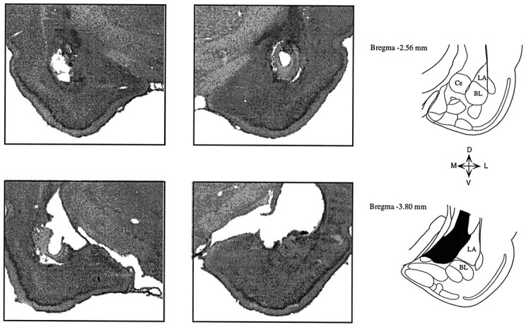
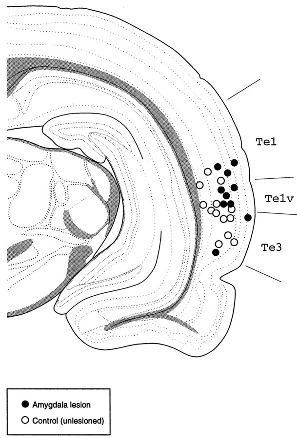
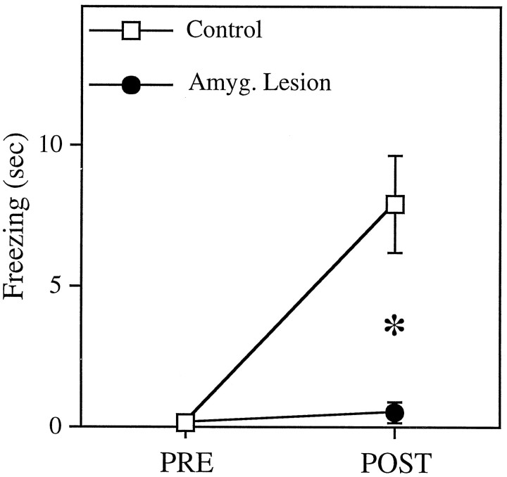
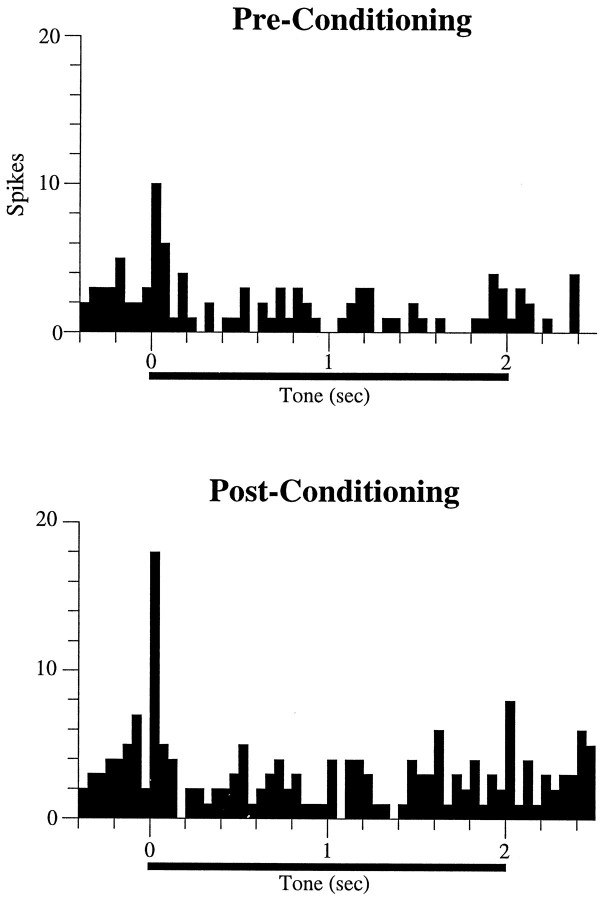
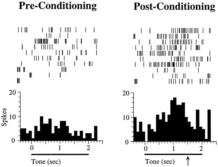
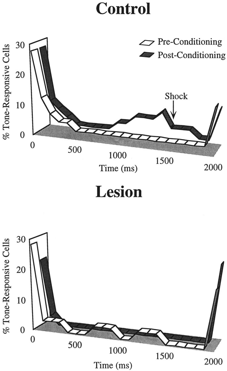
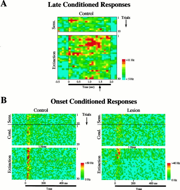
In auditory fear conditioning, pairing of a neutral acoustic conditioned stimulus (CS) with an aversive unconditioned stimulus (US) results in an enhancement of neural responses to the CS in the amygdala and auditory cortex. It is not clear, however, whether cortical plasticity governs neural changes in the amygdala or vice versa, or whether learning in these two structures is determined by independent processes. We examined this issue by recording single-cell activity in the auditory cortex (areas Te1, Te1v, and Te3) of freely behaving, amygdalectomized rats using a movable bundle of microwires. Amygdala damage did not affect short-latency (0-50 msec) tone responses, nor did it interfere with conditioning-induced increases of these onset responses. In contrast, lesions of the amygdala interfered with the development of late (500-1500 msec) conditioned tone responses that were not present before conditioning. Furthermore, whereas onset conditioned responses in the control group remained elevated after 30 extinction trials (presentation of CS alone), onset responses in lesioned animals returned to their preconditioning firing level after approximately 10 extinction trials. These results suggest that the amygdala enables the development of long-latency (US anticipatory) responses and prevents the extinction of short-latency onset responses to threatening stimuli. The findings further suggest that auditory cortex cells may participate differently in explicit and implicit memory networks.


Figures

Fig. 1.
Fig. 1.
Photomicrographs of coronal sections showing a typical bilateral amygdala lesion. Lesions were centered at the lateral and basolateral nuclei, and in some cases damage extended into the central nucleus. Damage to the caudate nucleus above the amygdala was variable from case to case. Ce, Central nucleus;LA, lateral nucleus; BL, basolateral nucleus.
Fig. 2.
Fig. 2.
Drawing of a coronal section, at ∼5 mm posterior to bregma, showing the electrode placements for control (open circles) and lesioned (filled circles) animals. Each circle corresponds to one electrode placement (each rat was implanted only once). Most placements were located in areas Te1v/Te3, which are part of the auditory association cortex in the rat. Te1, Te1v, Te3, Temporal cortex, areas 1–3. Adapted from Swanson (1992).
Fig. 3.
Fig. 3.
Freezing responses to the 20 sec tone conditioned stimulus (CS) before and after conditioning for control (n = 14) and lesion (n = 11) groups. After conditioning, control rats exhibited a significant increase in freezing to the CS (p < 0.001), whereas amygdala-lesioned animals showed no change.
Fig. 4.
Fig. 4.
Poststimulus time histograms showing the tone response of a cell in an amygdala-lesioned rat. This cell exhibited a short-latency (<50 msec) onset response before conditioning. As a result of conditioning, the magnitude of the onset response increased relative to the sensitization phase (Pre-Conditioning). Each histogram represents 10 trials; bin width is 50 msec.
Fig. 5.
Fig. 5.
Example of a cell in a control rat showing long-latency conditioned tone responses. Before conditioning, the cell did not respond to the tone. After conditioning, however, the cell developed a significant tone response, starting at ∼500 msec after tone onset and achieving its peak at 1500 msec after tone onset, which was the time of footshock onset during conditioning (indicated by thearrow). Shock was not delivered during these tests, because they occurred during extinction trials. Each histogram represents 10 trials; bin size is 100 msec. No cells showing these late conditioned responses were found in lesioned animals (see Results).
Fig. 6.
Fig. 6.
Ribbon plots showing the percentage of cells that were significantly tone responsive (see Materials and Methods) before (white) and after (gray) conditioning for the control and lesion groups. Although there were prominent onset and offset responses before and after conditioning in both groups, conditioning resulted in an increase in the number of cells in the control group showing late responses to the tone CS, starting at ∼900 msec after tone onset and reaching a peak at 1500 msec (time of shock onset during conditioning). This conditioning-induced late response was absent in lesioned animals. The duration of the tone was 2 sec; bin width is 100 msec.
Fig. 7.
Fig. 7.
A, Color-coded raster plot showing the average firing rate of cells in the control group that exhibited significant late tone responses (500–1500 msec after tone onset; see Results) after conditioning (n = 11). Thetop of the figure shows the sensitization trials (preconditioning) and the bottom shows extinction trials (the conditioning phase is not shown). Each row is one trial, and the bin size is 100 msec. Firing rates were averaged across cells and smoothed with a nearest-neighbor average. Note that there was no indication of late responses before conditioning, and that the long-latency conditioned response is maximal (red) just before the time of the shock onset during conditioning (indicated by the arrow), similar to the example shown in Figure 5.B, Color-coded raster plot showing the average firing rate of cells that develop short-latency (<50 msec) onset conditioned increases, for the control (n = 12) and lesion (n = 12) groups. Each row is one trial, showing the first 500 msec after tone onset; bin size is 10 msec. The top half shows the 10 sensitization and 20 conditioning trials, and the bottom half shows the 30 extinction trials. Animals were returned to their home cages for 1 hr after the end of conditioning and before the beginning of extinction. Note that in the control group the magnitude of onset responses after 30 extinction trials remained elevated relative to the response before conditioning (sensitization). In contrast, cells in lesioned animals returned to their preconditioning levels after 10 extinction trials.
Fig. 8.
Fig. 8.
Poststimulus time histograms showing the effects of extinction on short-latency (<50 msec) onset conditioning in cells from control (left) and lesioned (right) animals. The cell from the control animal developed onset conditioned increases after conditioning (extinction trials 1–10) that lasted through the entire extinction phase (30 trials). In contrast, the cell from the lesioned rat developed a short-lasting onset conditioned increase and returned to its preconditioning response level at the end of the extinction phase.
Fig. 9.
Fig. 9.
Average onset tone responses (0–50 msec) of cells that conditioned, from the control and lesion groups, plotted for the 10 sensitization trials (Pre Conditioning), the first 10 trials of extinction (Post Conditioning), and the last 10 trials of extinction (trials 20–30; Post Extinction), relative to the postconditioning tone response. In both groups, tone responses before conditioning were significantly smaller than after conditioning (p < 0.01). In the control group, the responses at the end of extinction did not differ from the postconditioning values (p> 0.1), whereas in the lesion group extinction trials resulted in a significant reduction of the tone responses (p < 0.005).

References

    1. Amaral DG. In: Memory: anatomical organization of candidate brain regions. Handbook of physiology. Section 1: The nervous system. Plum F, editor. American Physiological Society; Bethesda, MD: 1987. pp. 211–294.
    1. Amaral DG, Price JL, Pitkanen A, Carmichael ST. Anatomical organization of the primate amygdaloid complex. In: Aggleton JP, editor. The amygdala: neurobiological aspects of emotion, memory, and mental dysfunction. Wiley-Liss; New York: 1992. pp. 1–66.
    1. Armony JL, Servan-Schreiber D, Cohen JD, LeDoux JE. An anatomically constrained neural network model of fear conditioning. Behav Neurosci. 1995;109:246–257. - PubMed
    1. Armony JL, Servan-Schreiber D, Romanski LM, Cohen JD, LeDoux JE. Stimulus generalization of fear responses: effects of auditory cortex lesions in a computational model and in rats. Cereb Cortex. 1997a;7:157–165. - PubMed
    1. Armony JL, Servan-Schreiber D, Cohen JD, LeDoux JE. Computational modeling of emotion: explorations through the anatomy and physiology of fear conditioning. Trends Cognit Sci. 1997b;1:28–34. - PubMed

Publication types

LinkOut - more resources