. 1998 Apr 1;18(7):2788-99.
doi: 10.1523/JNEUROSCI.18-07-02788.1998.

In vivo modulation of interacting central pattern generators in lobster stomatogastric ganglion: influence of feeding and partial pressure of oxygen

Affiliations

In vivo modulation of interacting central pattern generators in lobster stomatogastric ganglion: influence of feeding and partial pressure of oxygen

S Clemens et al. J Neurosci. .

Abstract

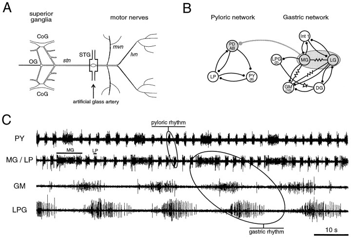
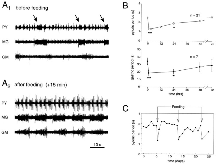
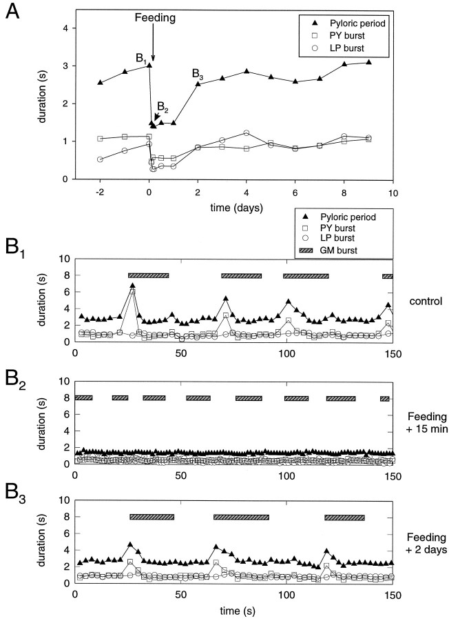
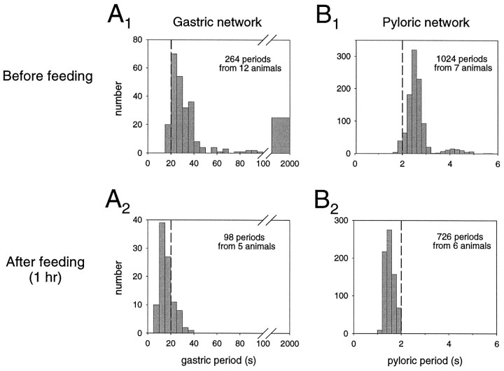
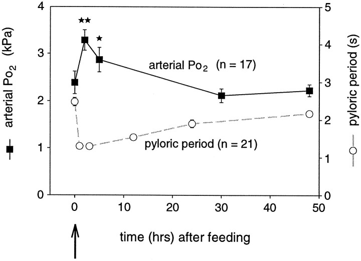
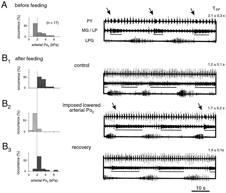
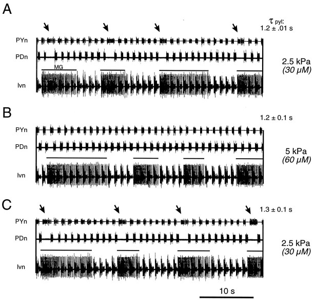
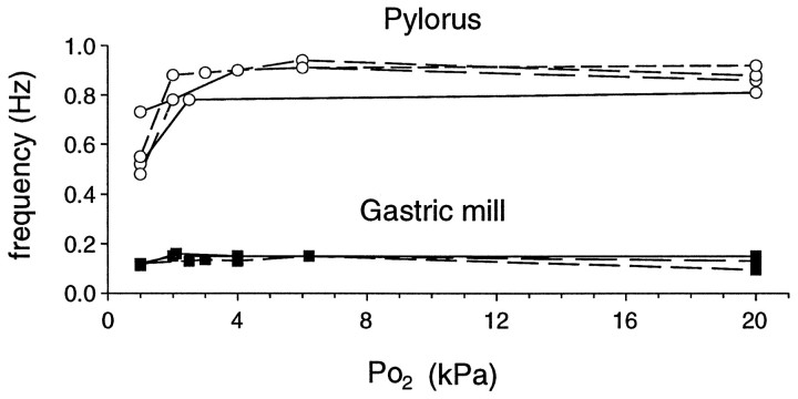
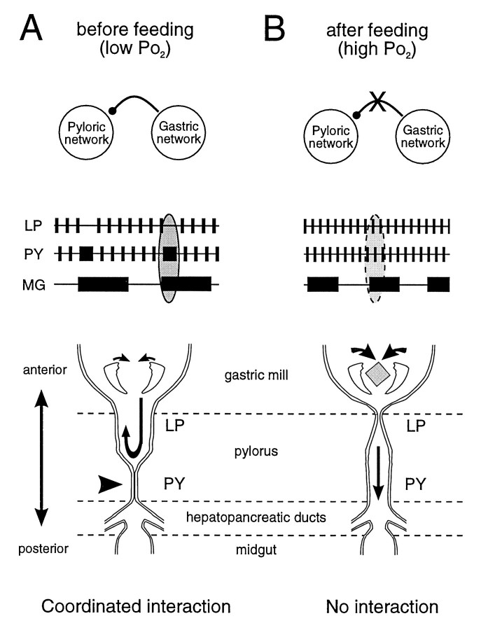
The stomatogastric ganglion (STG) of the European lobster Homarus gammarus contains two rhythm-generating networks (the gastric and pyloric circuits) that in resting, unfed animals produce two distinct, yet strongly interacting, motor patterns. By using simultaneous EMG recordings from the gastric and pyloric muscles in vivo, we found that after feeding, the gastropyloric interaction disappears as the two networks express accelerated motor rhythms. The return to control levels of network activity occurs progressively over the following 1-2 d and is associated with a gradual reappearance of the gastropyloric interaction. In parallel with this change in network activity is an alteration of oxygen levels in the blood. In resting, unfed animals, arterial partial pressure of oxygen (PO2) is most often between 1 and 2 kPa and then doubles within 1 hr after feeding, before returning to control values some 24 hr later. In vivo, experimental prevention of the arterial PO2 increase after feeding leads to a slowing of pyloric rhythmicity toward control values and a reappearance of the gastropyloric interaction, without apparent effect on gastric network operation. Using in vitro preparations of the stomatogastric nervous system and by changing oxygen levels uniquely at the level of the STG within the range observed in the intact animal, we were able to mimic most of the effects observed in vivo. Our data indicate that the gastropyloric interaction appears only during a "free run" mode of foregut activity and that the coordinated operation of multiple neural networks may be modulated by local changes in oxygenation.


Figures

Fig. 1.
Fig. 1.
Stomatogastric nervous system (STNS) of the European lobster H. gammarus. A, The dissected STNS in vitro. The stomatogastric ganglion (STG) receives descending input from the brain and other STNS ganglia via the single stomatogastric nerve (stn). For in vitro experiments, the STG was placed in an artificial glass artery to permit oxygenation changes at the level of the ganglion only. B, Synaptic wiring diagram of the pyloric and gastric networks. The pyloric network consists of 11 motor neurons and 1 interneuron (AB), and the gastric circuit is composed of 15 motor neurons and 1 interneuron (Int 1). An inhibitory synapse between the gastric neuronsMG/LG and the pyloric pacemaker groupPD/AB mediates an internetwork connection. Stick and ball symbols denote chemical inhibitory synapses, resistor symbols represent electrical connections, and diode indicates rectifying electrical coupling. C, Spontaneous activity of pyloric and gastric circuits in vivo. Simultaneous recording of the pyloric muscles innervated by LP neuron and PY neurons (pyloric rhythm) and of three gastric muscles innervated by MG neurons, GM neurons, and LPG neurons, respectively (gastric rhythm). Note that after the onset of each MG neuron burst the following PY neuron burst is substantially prolonged, which is caused by the inhibitory interaction between the gastric and pyloric networks. CoG, Commissural ganglion;OG, esophageal ganglion; lvn, lateroventricular nerve; mvn, medioventricular nerve;PD, pyloric dilator; AB, anterior burster; LP, lateral pyloric constrictor;PY, pyloric constrictor; Int 1, interneuron 1; LPG, lateral posterior gastric;MG, medial gastric; LG, lateral gastric;GM, gastric mill; DG; dorsal gastric.
Fig. 2.
Fig. 2.
Effects of feeding on motor expression of the STG networks. A1, EMG recordings from pyloric (innervated by PY neurons) and gastric muscles (innervated by MG and GM neurons) before feeding. Pyloric and gastric networks oscillate at periods of 2.5 sec and 30–40 sec, respectively. Note PY neuron burst prolongation after onset of each MG neuron burst (arrows). A2, EMG recordings from the same animal after feeding. Both networks oscillate at higher frequencies, and the gastropyloric interaction is no longer evident. Note that the fraction of each gastric cycle during which MG and GM neurons are active is increased considerably. B, Pooled data showing the effects of feeding on pyloric (top) and gastric (bottom) network activity. Each point is the mean period ± SE of 5 min samples. Significant differences in motor expression persist for at least 24 hr after ingestion. **p < 0.01; *p < 0.05; Mann–Whitney test. C, Repetitive feeding (arrows) elicits reproducible accelerations of pyloric activity in a single animal during a 4 week recording period. Each feeding stimulus accelerates pyloric activity by ∼50%, albeit with varying recovery slopes.
Fig. 3.
Fig. 3.
Evolution of pyloric activity and gastropyloric interaction after feeding. A, Typical time courses of mean pyloric periods and LP and PY neuron burst duration after feeding. All three parameters are reduced abruptly and then recover gradually over the ensuing 2 d. B, Cycle-by-cycle analysis of EMG samples in the same experiment before feeding (B1), 15 min after feeding (B2), and 2 d after feeding (B3). B1, Before feeding, pyloric periods (black triangles) are transiently prolonged after the onset of each recorded GM neuron burst (seebar). This is associated with a considerable prolongation of PY neuron (square symbol) burst duration only. B2, Fifteen minutes after feeding, both networks oscillate at higher frequencies, LP neuron (open circle) and PY neuron burst durations decrease, and the pyloric perturbation has completely vanished.B3, Two days after feeding, the rhythmic gastropyloric interaction reappears as the activity of the two networks returns toward control values.
Fig. 4.
Fig. 4.
Distribution histograms of gastric and pyloric periods before and after feeding. A, Gastric network.A1, Before feeding, gastric periods are widely varied, mostly occurring between 20 and 40 sec. Note that periods >100 sec were considered as pauses. A2, After feeding, gastric periods decreased to 10–20 sec without pauses.B, Pyloric circuit. B1, Before feeding, pyloric periods display a bimodal distribution at 2.4–2.8 sec and at 3.8–4.4 sec. The latter mode corresponds to the rhythmic perturbations by gastric activity. B2, After feeding, the second mode disappears with pyloric periods now occurring mostly between 1.4 and 1.8 sec.
Fig. 5.
Fig. 5.
Time course of mean arterial Po2 after feeding (filled squares, solid line). For comparison, the postprandial time course of pyloric period after feeding, as shown in Figure2B, is also plotted (open circles, dashed line). After feeding, as pyloric period decreases, arterial Po2 increases significantly and then both parameters return gradually toward control values. **p < 0.01; *p < 0.05; Mann–Whitney test.
Fig. 6.
Fig. 6.
Transient hypoxia partially reverses postprandial changes in vivo. A, Distribution histogram of arterial Po2 in resting unfed animals and corresponding pyloric and gastric motor patterns. Po2 lies mostly in the range of 1–2 kPa. Characteristically, cycle periods are long, and each gastric MG neuron burst (underlined) is associated with a pyloric network perturbation (arrows). τpylpyloric cycle period. B, Effects of imposed lowered arterial Po2 after feeding.B1, Under control conditions, after feeding, the normoxic arterial Po2 increases along with the rate of pyloric and gastric cycling, and the internetwork interaction disappears. B2, Transient suppression of postprandial Po2increase (by lowering water Po2) reverses pyloric (but not gastric) acceleration, and the internetwork interaction is restored (arrows).B3, These effects are reversible.
Fig. 7.
Fig. 7.
Selective action of Po2 on gastropyloric interaction in vitro. A, During superfusion of the STG alone with equilibrated saline at Po2 = 2.5 kPa, the spontaneously active gastric and pyloric networks express an interaction (arrows) similar to that seen in resting and unfed animals. Each onset of a gastric MG neuron burst (solid line above lvn trace) is followed by a single perturbation in the pyloric network cycle.B, Superfusing the STG with saline oxygenated at 5 kPa leads to a disappearance of the internetwork interaction, without affecting pyloric or gastric cycle frequency in this Po2 range. C, This specific action is reversed on return to control conditions. τpyl, Pyloric cycle period.
Fig. 8.
Fig. 8.
Differential effects of Po2 on pyloric and gastric networks in vitro. Pyloric activity is considerably influenced by different oxygenation levels only in the physiological range of 1–3 kPa. By contrast, gastric cycling remains unaffected (n = 4 preparations).
Fig. 9.
Fig. 9.
Model of proposed changes in foregut function before and after feeding. A, Before feeding, at low Po2, the gastric and pyloric networks cycle slowly and express an interaction in which PY and MG neuron bursts are coordinated. During the sustained contraction of the posterior part of the pylorus attributable to a longer-lasting drive from the PY neurons, the anterior region (controlled by the LP neuron) will remain open. This may allow a pumping action of the gastric teeth to transfer digestive enzymes, present in the pylorus, toward the stomach and esophagus. B, After feeding, at higher Po2, the two networks now perform their separate behavioral tasks (masticatory function of the gastric mill and filtering function of the pylorus) without a coordinating interaction. Note that the switch from one functional state to the other is under the control of local Po2 changes at the STG level.

References

    1. Aiken DE. Proecdysis, setal development, and molt predictions in the American lobster (Homarus americanus). J Fish Res Board Can. 1973;30:1337–1344.
    1. Barker PL, Gibson R. Observations on the feeding mechanism, structure of the gut, and digestive physiology of the European lobster Homarus gammarus. J Exp Mar Biol Ecol. 1977;26:297–324.
    1. Bernasconi P, Kohl J. Analysis of co-ordination between breathing and exercise rhythms in man. J Physiol (Lond) 1993;471:693–706. - PMC - PubMed
    1. Bredt DS, Snyder SH. Nitric oxide, a novel neuronal messenger. Neuron. 1992;8:3–11. - PubMed
    1. Bredt DS, Hwang PM, Snyder SH. Localization of nitric oxide synthase indicating a neural role for nitric oxide. Nature. 1990;347:768–770. - PubMed

Publication types

LinkOut - more resources