. 1998 Feb 1;506 ( Pt 3)(Pt 3):731-44.
doi: 10.1111/j.1469-7793.1998.731bv.x.

Glycinergic synaptic inputs to bipolar cells in the salamander retina

Affiliations

Glycinergic synaptic inputs to bipolar cells in the salamander retina

B R Maple et al. J Physiol. .

Abstract

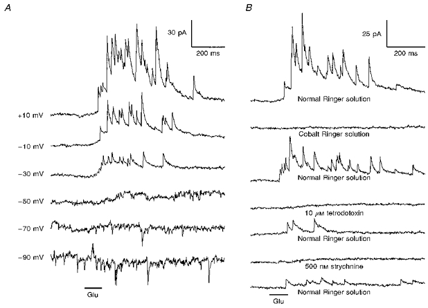
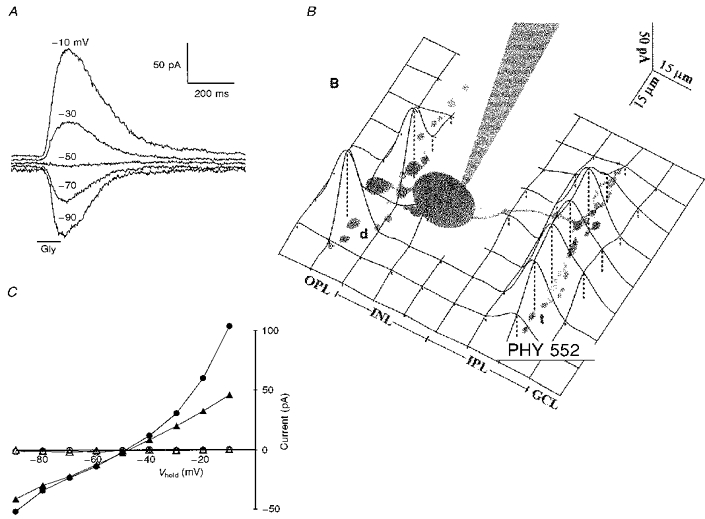
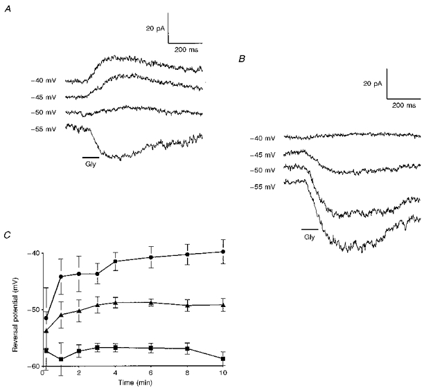
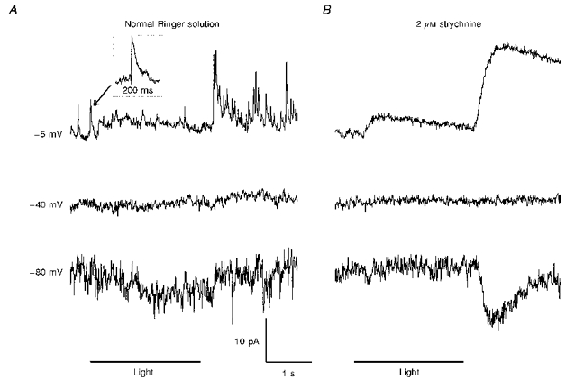
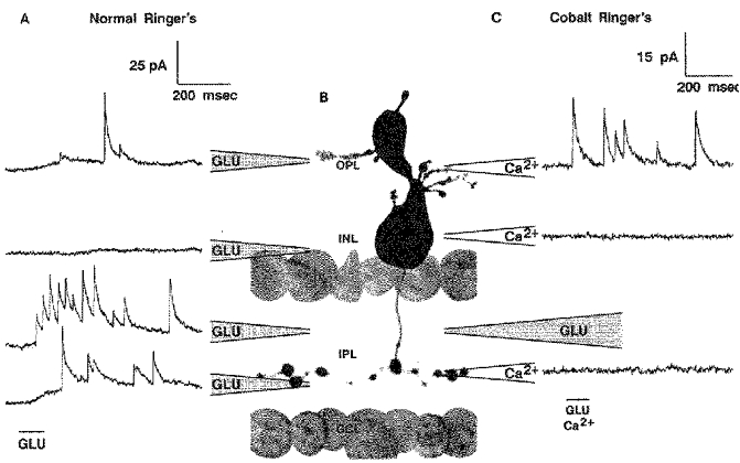
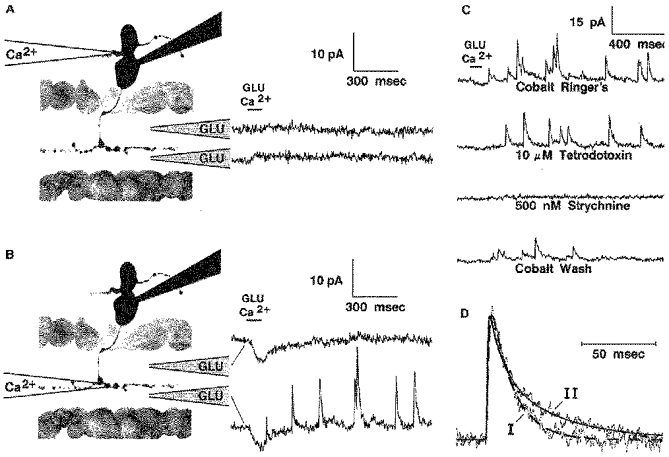
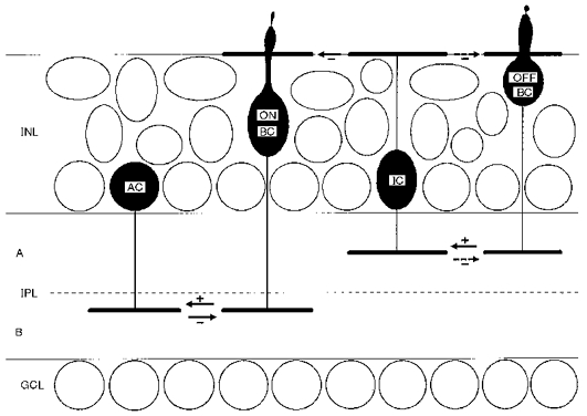
1. Glycine activated strychnine-sensitive chloride conductances at both the dendrites and the axonal telodendria of most bipolar cells in the salamander retina. 2. The chloride equilibrium potential of bipolar cells was found to be negative to -50 mV, indicating that glycinergic synapses on bipolar cells are inhibitory. 3. Some bipolar cells exhibited discrete, strychnine-sensitive, chloride-mediated inhibitory postsynaptic currents (IPSCs). These were elicited by focal application of glutamate at the inner plexiform layer (IPL). Glycinergic synapses were localized using simultaneous focal application of calcium to retinal slices bathed in calcium-free media. Both dendritic and telodendritic glycinergic IPSCs were observed. 4. The decay of the telodendritic IPSCs was well fitted by a single exponential with a time constant of 17.7 +/- 8.7 ms. Similar kinetics were observed for dendritic IPSCs in some cells, but in one class of on-centre bipolar cell the decay of the dendritic IPSCs was better fitted by a sum of two exponentials with time constants 9.9 +/- 4.3 and 51.3 +/- 24.3 ms. 5. The dendritic IPSCs were best driven by application of glutamate at the distal IPL (the off sublamina), while the telodendritic IPSCs were driven best by application near the telodendria. These results suggest that bipolar cell dendrites receive inhibitory glycinergic inputs from interplexiform cells that are excited by off-centre bipolar cells, whereas bipolar cell telodendria receive glycinergic amacrine cell inputs that are antagonistic to the photoreceptor inputs. 6. Both inputs could be elicited in the presence of tetrodotoxin (TTX), but the dendritic IPSCs were sometimes abolished by TTX, suggesting that sodium-dependent spikes play an important role in the transmission of interplexiform cell signals to the outer plexiform layer.


Figures

Figure 4
Figure 4. Strychnine-sensitive IPSCs elicited by stimulation with glutamate
A, responses of an on-centre (0.7 IU) bipolar cell to focal pressure ejection of 100 μM glutamate (Glu) at the distal region of the IPL. The holding potential for each record is indicated on the left. The IPSCs reversed near ECl (set to -50 mV in this experiment). B, the IPSCs were reversibly abolished by substitution of Co2+ for Ca2+, and by the addition of 10 μM tetrodotoxin or 500 nM strychnine to the bath. The holding potential was -10 mV.
Figure 1
Figure 1. Voltage clamp responses of bipolar cells to iontophoretic application of glycine
A, responses of a bipolar cell to application of glycine (Gly) at the axonal telodendria. B, a spatial profile for glycine sensitivity generated by applying glycine to many sites with the bipolar cell held at -10 mV. The height of the surface (indicated by dashed lines) gives the magnitude of the current response at each location (d, dendrites; t, telodendria; OPL, outer plexiform layer; IPL, inner plexiform layer; INL, inner nuclear layer; GCL, ganglion cell layer). C, the mean current response as a function of holding potential for dendritic application (○, •) and telodendritic application (▵, ▴) of glycine to 10 cells (5 on-centre and 5 off-centre). The filled symbols indicate responses obtained in Co2+ Ringer solution. The open symbols indicate responses for the same cells in Co2+ Ringer solution plus 1 μM strychnine. (For 5 cells glycine was applied first at the dendrites, then at the telodendria, then again at the same telodendritic site after strychnine had been applied. For the other five cells the order was reversed and strychnine was applied while the iontophoretic pipette was positioned at the dendrites.) The chloride equilibrium potential was set to -50 mV in these experiments.
Figure 2
Figure 2. Estimation of the normal physiological ECl for bipolar cells
A, voltage clamp responses of a bipolar cell to pressure ejection of 100 μM glycine (Gly) at the OPL immediately after the voltage clamp was established. B, responses from the same cell after 10 min of internal perfusion. (ECl was set to -40 mV in this experiment). C, the mean reversal potential as a function of time (obtained by interpolation between the smallest positive and negative responses) for ECl= -40 mV (•), ECl= -50 mV (▴), and ECl= -60 mV (▪). Data from 8 on-centre cells, 3 off-centre cells, and 4 axotomized or multistratified cells.
Figure 3
Figure 3. The effect of light on the frequency of spontaneous strychnine-sensitive IPSCs
A, responses of an on-centre (0.7 IU) bipolar cell to diffuse illumination. Discrete IPSCs, suppressed by light and strongly elicited by light off, reversed near ECl (set to -40 mV in this experiment). B, 2 μM strychnine abolished the discrete IPSCs, leaving an enhanced on-off inhibition that was not obviously composed of discrete events.
Figure 5
Figure 5. Characterization of dendritic glycinergic IPSCs
A, a spatial profile for the glutamate (GLU) response of the cell of Fig. 4. The IPSCs were best elicited by application of glutamate at the distal IPL, although a weaker response was also isolated to the OPL. B, a photograph of another bipolar cell with morphology and glutamate response profile similar to that of the one in A. C, responses of the cell depicted in B to application of glutamate at the distal IPL. In this case the cell was bathed in Co2+ Ringer solution and Ca2+ Ringer solution was simultaneously applied focally to different regions of the slice. IPSCs were elicited only when calcium was applied at the OPL. The holding potential was -10 mV for all traces.
Figure 6
Figure 6. Characterization of telodendritic glycinergic IPSCs
Responses are shown for another on-centre bipolar cell stimulated by simultaneous focal application of glutamate (Glu) and calcium in a Co2+ Ringer solution bath. In this case no IPSCs were elicited when calcium was applied to the dendrites (A). IPSCs were observed when calcium was applied to the telodendria, but only when glutamate was also applied at the proximal IPL (B). The telodendritic IPSCs were relatively unaffected by 10 μM TTX, but were reversibly abolished by 500 nM strychnine (C). In D the time course of a telodendritic IPSC from this cell (I) is compared with that of a dendritic IPSC (II) from the cell of Fig. 5B. The broken line gives the best fit of the telodendritic IPSC by eqn (1) with τ1= 1.2 ms and τ2= 15.1 ms. The continuous line gives the best fit of the dendritic IPSC to eqn (2) with τ1= 0.39; τ2= 9.4 and τ3= 40.5 ms; C2= 0.54. The amplitudes of the IPSCs were normalized for purposes of comparison.
Figure 7
Figure 7. A model for the circuitry of glycinergic inputs to centrally-ramifying on-centre bipolar cells
The border between sublaminae A and B is indicated by the dashed line. Excitatory synapses are indicated by arrows with a (+) sign. Inhibitory (chloride-mediated) synapses are indicated by arrows with (-) signs. AC, amacrine cell; BC, bipolar cell; GCL, ganglion cell layer; IC, interplexiform cell; INL, inner nuclear layer; IPL, inner plexiform layer.

Similar articles

Cited by

References

    1. Attwell D, Mobbs P, Tessier-Lavigne M, Wilson M. Neurotransmitter-induced currents in retinal bipolar cells of the axolotl, Ambystoma mexicanum. The Journal of Physiology. 1987;387:125–161. - PMC - PubMed
    1. Dixon DB, Copenhagen DR. Two types of glutamate receptors differentially excite amacrine cells in the tiger salamander retina. The Journal of Physiology. 1992;449:589–606. - PMC - PubMed
    1. Famiglietti EV, Jr, Kaneko A, Tachibana M. Neuronal architecture of on and off pathways to ganglion cells in carp retina. Science. 1977;198:1267–1269. - PubMed
    1. Famiglietti EV, Jr, Kolb H. Structural basis for ON-and OFF-center responses in retinal ganglion cells. Science. 1976;194:193–195. - PubMed
    1. Gao F, Yang J-H, Wu SM. Miniature inhibitory postsynaptic currents in retinal ganglion cells. Investigative Ophthalmology and Visual Science. 1997;38:S51. abstract 242.

Publication types

MeSH terms

LinkOut - more resources