Case Reports
. 1997 Nov;81(11):980-3.
doi: 10.1136/bjo.81.11.980.

Clinical experiences of infectious scleral ulceration: a complication of pterygium operation

Affiliations
Case Reports

Clinical experiences of infectious scleral ulceration: a complication of pterygium operation

C P Lin et al. Br J Ophthalmol. 1997 Nov.

Abstract

Aims: To report the special clinical manifestations and determine the appropriate management of infectious scleral ulceration.

Methods: A retrospective study was performed on 30 eyes with infectious scleral ulceration. Information was recorded on patients' age, onset and course of disease, pathogenic organism, clinical presentations, methods of diagnosis, treatment, and outcome.

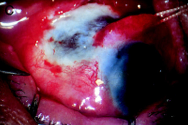
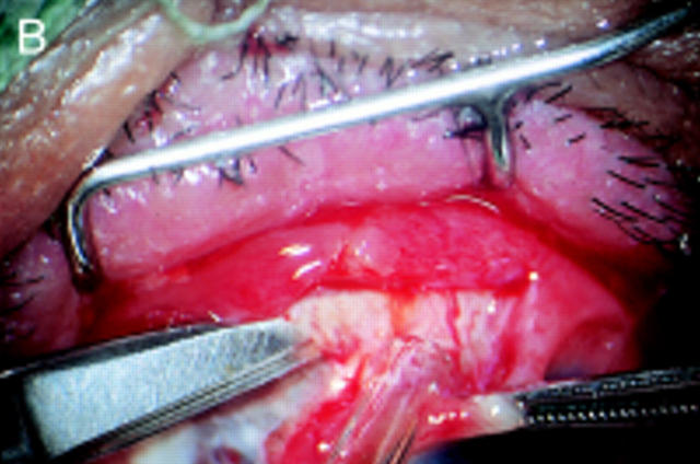
Results: 10 cases (33.3%) were accompanied by corneal involvement. Subconjunctival abscess was noted in 16 cases (53.3%). 17 cases (56.7%) gave positive results of pathogen culture and all were Pseudomonas aeruginosa. Two cases had combined bacterial infections and one case was complicated by fungal infection. A total of 26 cases had surgical debridement in this series. Extensive involvement of the sclera with the presence of a 'tunnel lesion' or a 'satellite subconjunctival abscess' were found during debridement. All of the eyeballs involved were salvaged except one.

Conclusion: The results of this study were contrary to the poor prognosis presented in previous reports. Early and repetitive surgical debridement is believed to be mandatory in the intractable cases to shorten the admission period and to save these eyes.


Figures

Figure 1
Figure 1
(A) The scleral ulceration was located at 9 o'clock position. Severe congestion and subconjunctival abscess were noted at 10 o'clock position under the biomicroscope. (B) After the surgical debridement, the scleral wound was left open. In comparison with (A) the involved area was more extensive. (C) Ten days later, the debrided area was healing.
Figure 2
Figure 2
(A) During the surgical debridement, a tunnel lesion was detected with spring scissors easily entering intrastromal space. (B) Removal of the necrotic scleral tissue with spring scissors under continuous irrigation and aspiration. (C) The area involved was much larger than the visible lesion before debridement.
Figure 2
Figure 2
(A) During the surgical debridement, a tunnel lesion was detected with spring scissors easily entering intrastromal space. (B) Removal of the necrotic scleral tissue with spring scissors under continuous irrigation and aspiration. (C) The area involved was much larger than the visible lesion before debridement.

References

    1. Arch Ophthalmol. 1980 Feb;98(2):341-5 - PubMed
    1. Infect Immun. 1978 Feb;19(2):630-48 - PubMed
    1. Br J Ophthalmol. 1980 Jul;64(7):496-505 - PubMed
    1. Br J Ophthalmol. 1980 Sep;64(9):676-9 - PubMed
    1. Am J Ophthalmol. 1981 Jun;91(6):706-10 - PubMed

Publication types