

U.S. case report of cerebral phaeohyphomycosis caused by Ramichloridium obovoideum (R. mackenziei): criteria for identification, therapy, and review of other known dematiaceous neurotropic taxa
- PMID: 9508300
- PMCID: PMC104613
- DOI: 10.1128/JCM.36.3.708-715.1998
U.S. case report of cerebral phaeohyphomycosis caused by Ramichloridium obovoideum (R. mackenziei): criteria for identification, therapy, and review of other known dematiaceous neurotropic taxa
Abstract
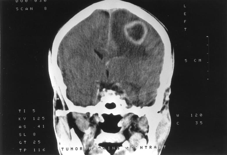
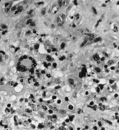
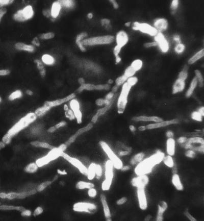
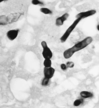
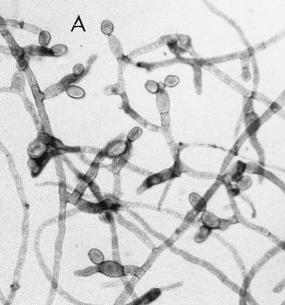
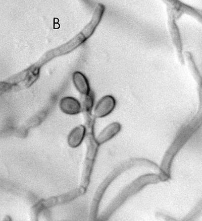
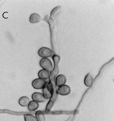
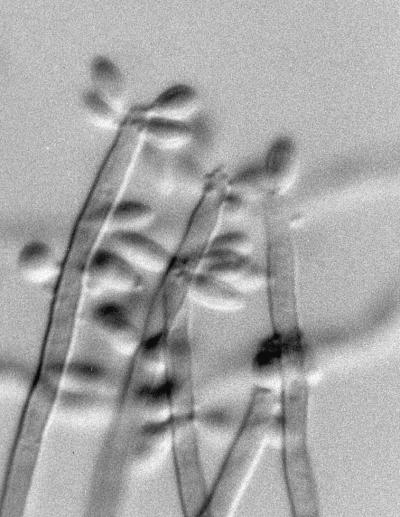
We report a case of cerebral phaeohyphomycosis in a 36-year-old male caused by the neurotropic fungus Ramichloridium obovoideum (Matushima) de Hoog 1977 (Ramichloridium mackenziei Campbell et Al-Hedaithy 1993). This man resided in the Middle East, where the fungus appears to be endemic and, possibly, geographically restricted, since all previous reports of brain abscesses due to this organism have been for patients indigenous to this area. As a servant of the Saudi Arabian royal family, he appeared in the United States seeking treatment for chronic weight loss, fatigue, decreased memory, and a more recent 2-week history of right-hand weakness which worsened to involve the entire right upper extremity. On the day prior to his admission, he had a focal motor seizure with rotation of the head and eyes to the right, followed by secondary generalization. A computerized tomogram showed a ring-enhancing hypodense lesion in the left parietal subcortical region with associated edema and mass effect. Diagnosis of a fungal etiology was made following a parietal craniotomy and excisional biopsy by observation of septate, dematiaceous hyphal elements 2 to 3 microm in width on hematoxylin-and-eosin-stained sections from within areas of inflammation and necrosis. Culture of the excised material grew out a dematiaceous mould which was subsequently identified as R. obovoideum. At two months postsurgery and with a regimen of 200 mg of itraconazole twice a day, the patient was doing well and returned to Saudi Arabia. His condition subsequently deteriorated, however, and following a 7-month course of itraconzole, he expired. We use this case to alert clinicians and personnel in clinical mycology laboratories of the pathogenicity of this organism and its potential occurrence in patients with central nervous system signs and symptoms who have resided in the Middle East and to review and/or compare R. obovoideum with other neurotropic, dematiaceous taxa and similar nonneurotropic, dematiaceous species.
Figures
References
-
- Al-Hedaithy, S. S. A., Z. A. B. Jamjoom, and E. S. Saeed. 1988. Cerebral phaeohyphomycosis caused by Fonsecaea pedrosoi in Saudi Arabia. Acta Pathol. Microbiol. Scand. 96(Suppl. 3):94–100. - PubMed
-
- Ayadi A, Huerre M-R, de Bievre C. Phaeohyphomycosis caused by Veronea botryosa. Lancet. 1995;346:1703–1704. - PubMed
-
- Bennett J E, Bonner H, Jennings A E, Lopez R I. Chronic meningitis caused by Cladosporium trichoides. Am J Clin Pathol. 1973;59:398–407. - PubMed
-
- Biggs P J, Allen R L, Powers M M, Holley H P. Phaeohyphomycosis complicating compound skull fracture. Surg Neurol. 1986;25:393–396. - PubMed
Publication types
MeSH terms
Substances
LinkOut - more resources
Full Text Sources
Medical
Research Materials
