

Review
doi: 10.1083/jcb.140.6.1281.
Leading the procession: new insights into kinesin motors
Affiliations
- PMID: 9508762
- PMCID: PMC2132666
- DOI: 10.1083/jcb.140.6.1281
Item in Clipboard
Review
Leading the procession: new insights into kinesin motors
J Cell Biol.
.
No abstract available
Figures
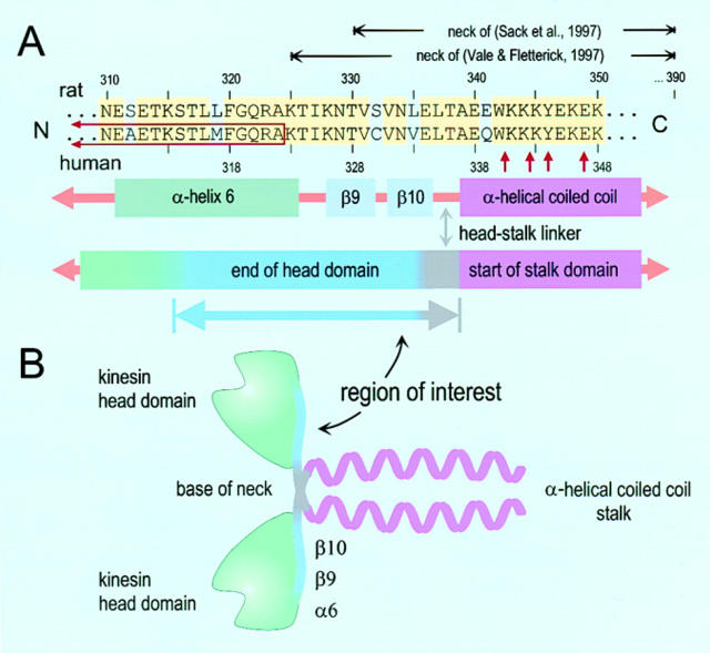
Sequence alignment for a portion of the kinesin heavy chain and corresponding elements in the kinesin structure. (A) Alignment of the rat (top) and human (bottom) sequences spanning the region ∼310–350 aa. Identical amino acids are colored (yellow). The numbering scheme for rat and human sequences differs by two. Neck regions, as defined by Sack et al. (21) and Vale and Fletterick (27), are indicated (horizontal black arrows). The sequence replaced in the ncd-kinesin chimera of Case et al. (6) is indicated on the left (red open box, left), as well as the sites of various mutations introduced by Romberg et al. (19) (vertical red arrows, right). Immediately under the alignment are structural elements of the dimer: α-helix 6 (olive), β-strands 9 and 10 (lavender), and the start of the coiled-coil (violet). Below these are the structural domains, consisting of the head (green, COOH terminus blue), head-stalk linker (gray), and initial portion of the stalk (violet). A new “region of interest” begins in α-helix 6 and continues until the start of the stalk (see text). (B) The corresponding structural domains are shown mapped onto a cartoon of the kinesin dimer, using the same coloring scheme.
References
-
- Alberts B. The cell as a collection of protein machines: preparing the next generation of molecular biologists. Cell. 1998;92:291–294. - PubMed
-
- Berliner E, Young EC, Anderson K, Mahtani HK, Gelles J. Failure of single-headed kinesin to track parallel to microtubule protofilaments. Nature. 1995;373:718–721. - PubMed
-
- Block SM. Fifty ways to love your lever: myosin motors. Cell. 1996;87:151–157. - PubMed
-
- Block, S.M. 1998. Kinesin molecular mechanics: what gives? Cell. In press.
-
- Block SM, Goldstein LSB, Schnapp BJ. Bead movement by single kinesin molecules studied with optical tweezers. Nature. 1990;348:348–352. - PubMed
Publication types
MeSH terms
Substances
LinkOut - more resources
Full Text Sources
Molecular Biology Databases
