. 1998 Mar 23;140(6):1285-95.
doi: 10.1083/jcb.140.6.1285.

Replicon clusters are stable units of chromosome structure: evidence that nuclear organization contributes to the efficient activation and propagation of S phase in human cells

Affiliations

Replicon clusters are stable units of chromosome structure: evidence that nuclear organization contributes to the efficient activation and propagation of S phase in human cells

D A Jackson et al. J Cell Biol. .

Abstract

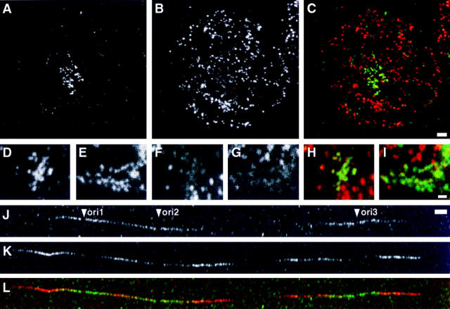
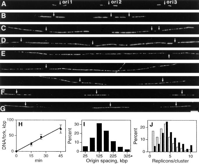
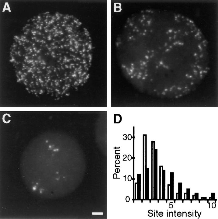
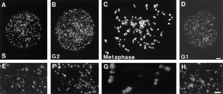
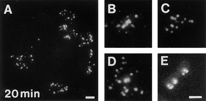
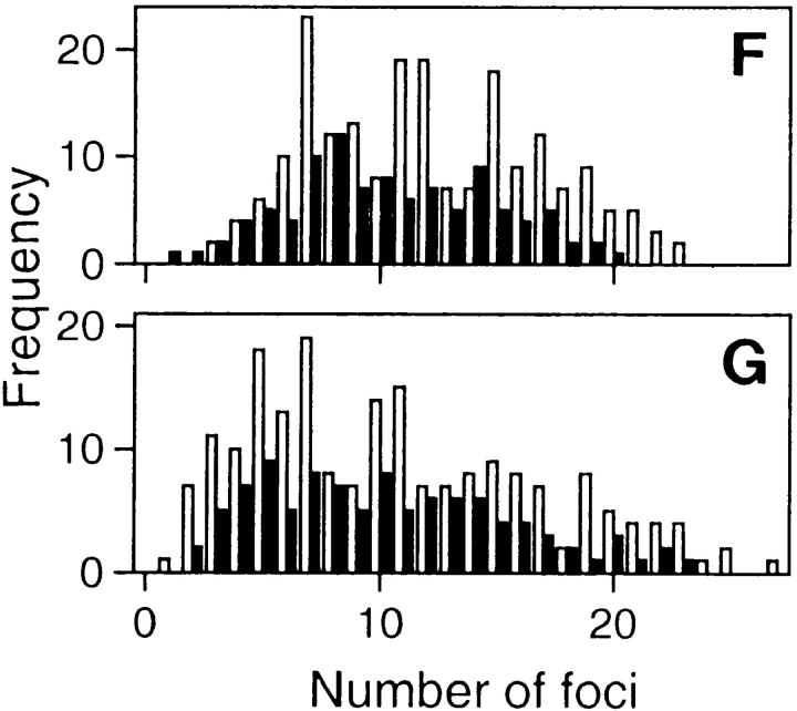
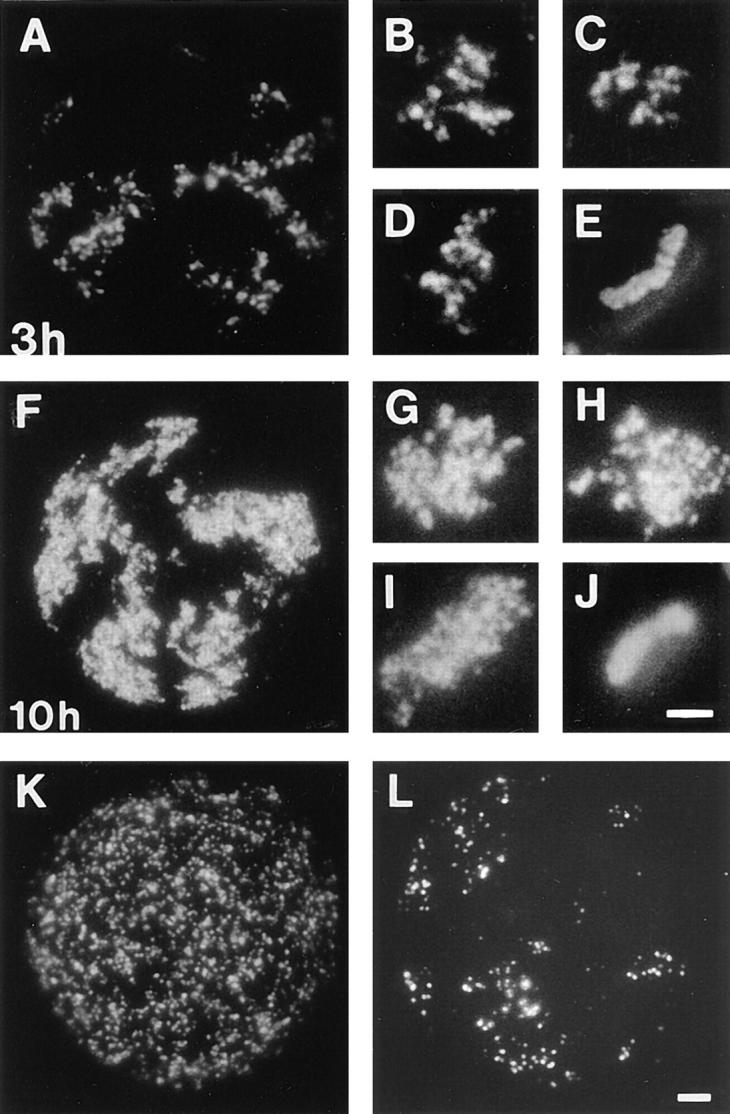
In proliferating cells, DNA synthesis must be performed with extreme precision. We show that groups of replicons, labeled together as replicon clusters, form stable units of chromosome structure. HeLa cells were labeled with 5-bromodeoxyuridine (BrdU) at different times of S phase. At the onset of S phase, clusters of replicons were activated in each of approximately 750 replication sites. The majority of these replication "foci" were shown to be individual replicon clusters that remained together, as stable cohorts, throughout the following 15 cell cycles. In individual cells, the same replication foci were labeled with BrdU and 5-iododeoxyuridine at the beginning of different cell cycles. In DNA fibers, 95% of replicons in replicon clusters that were labeled at the beginning of one S phase were also labeled at the beginning of the next. This shows that a subset of origins are activated both reliably and efficiently in different cycles. The majority of replication forks activated at the onset of S phase terminated 45-60 min later. During this interval, secondary replicon clusters became active. However, while the activation of early replicons is synchronized at the onset of S phase, different secondary clusters were activated at different times. Nevertheless, replication foci pulse labeled during any short interval of S phase were stable for many cell cycles. We propose that the coordinated replication of related groups of replicons, that form stable replicon clusters, contributes to the efficient activation and propagation of S phase in mammalian cells.


Figures

Figure 6
Figure 6
Early replicons are activated efficiently in different cell cycles. Cells synchronized at the beginning of S phase were grown for 20 min in medium containing BrdU, and then 4 d in medium, synchronized a second time, and grown for 20 min in medium containing IdU. Sites containing BrdU (A, D, and E) and IdU (B, F, and G) were indirectly immunolabeled. CLSM images showed foci containing Br-DNA (green) to colocalize with IdU-labeled (red) sites (C, H, and I; overlaps between A, D, or E, and B, F, or G show yellow in merge). Synchronized cells were also grown in medium supplemented with BrdU 5–20 min from the beginning of one S phase and IdU 10–30 min from the beginning of the next, after growing for 20 h in medium and synchronizing a second time (J). Spread DNA fibers were prepared and sites containing BrdU (J) and IdU (K) incorporation indirectly immunolabeled with FITC and Cy-3, respectively. Clusters of two to four replicons with at least one replicon labeled with both BrdU and IdU were collected; a typical example with three replicons (ori1–3) that are active in both cycles is shown (J–L). In 54 clusters with 126 replicons 120 were labeled with both analogues. Bars: (A–C) 2.5 μm; (D–I) 1 μm; (J–L) 5 μm.
Figure 4
Figure 4
Replication foci contain multiple replicons. Cells synchronized at the beginning of S phase were grown for 5 min in medium, and then 10 (A), 20 (B), 40 (C), 60 (D), or 120 min (E–G) in medium containing BrdU. Spread DNA fibers were prepared and sites of BrdU incorporation indirectly immunolabeled with Cy-3 (A–G). Each panel (A–G) shows a single- labeled DNA fiber with three origins of replication (ori1–3, small arrows from left to right). In A–E, replication is assumed to have initiated at the center of a short, unlabeled region flanked by symmetrical growing forks. Replicons activated within S phase (E and F) are uniformly labeled, their origins are assumed to lie centrally. In E, three panels show two early origins (ori1, and 2) apparently adjacent to one that fired much later (ori3); for orientation, ori1 is indicated in both top and center panels. The rate of replication fork movement was established (H). For the times shown, 50 replicons (A–C) were measured and the average lengths (± SD) of 100 forks determined. The separation of 250 adjacent origins activated at the onset of S phase (I) and number of replicons activated in 100 clusters (J) are shown. In J, open bars show the distribution of clusters and closed bars the distribution of DNA. Bar, 5 μm.
Figure 7
Figure 7
Early replication foci and transcription sites have common structural features. Synchronized cells encapsulated in agarose microbeads were grown in medium containing BrdU for 20 min either at the beginning of S phase (A) or 3 h later (B). Samples were extracted with 2 M NaCl and Br-DNA indirectly immunolabeled with FITC. G1 phase cells were grown in medium supplemented with BrU for 5 min and transcription sites labeled in the same way (C). Hypertonic extraction removes histones and spreads ∼60% DNA into a “halo” that surrounds the remnant nuclear matrix (D; DAPI staining of C). Bar, 2 μm.
Figure 3
Figure 3
The fluorescence intensities of replication foci. Cells synchronized at the beginning of S phase were grown for 20 min in medium containing BrdU, and then 20 h or 5 d in medium. Sites of BrdU incorporation were indirectly immunolabeled with FITC and images recorded using a CCD camera. A shows a typical cell in the first G1 phase after labeling and (B and C) two cells that indicate the range of labeling 5 d later. Raw images were collected, noise removed, and the intensities of individual foci were determined (D). 621 sites (D, open bars) chosen from random areas of six cells like A, and 430 sites (D, closed bars) all foci from seven cells like B and C were measured. Bar, 2.5 μm.
Figure 1
Figure 1
Sites of DNA synthesis in synchronized HeLa cells. Cells synchronized at the beginning of S phase were grown for 20 min in medium containing BrdU. Sites of incorporation were indirectly immunolabeled with FITC immediately (A and E), and 10 (B and F), 15 (C and G), and 20 h (D and H) later. All the labeled sites of typical cells (A–D), and details from different cells (E–H) are shown. Arrowheads (C) mark the ends of a TOTO- 3–stained chromosome that labeled poorly. Based on size, it is probably the predominantly late-replicating, inactive X chromosome. Double arrowheads in F highlight daughter replication foci seen in G2 phase of the cell cycle; typical low (small arrowheads) and high (large arrowheads) intensity sites are indicated. Bars: (A–D) 2 μm; (E–F) 1 μm.
Figure 2
Figure 2
Replicon clusters are stable over many cell generations. Cells synchronized at the beginning of S phase were grown for 20 min in medium containing BrdU and then 5 or 10 d in medium. Sites of BrdU incorporation were indirectly immunolabeled with FITC 5 (A) or 10 d (B–E) later. A shows a single cell with many clusters of labeled foci and (B–D) isolated clusters of labeled foci, the only labeled sites within three different cells. Foci are scattered along metaphase chromosomes in the same preparations (E); double exposure shows that foci are restricted to only one chromatid. The foci of 200 discrete clusters immunolabeled 5–10 d after incorporating BrdU in cells at the beginning of S phase (F), or unsynchronized cells (G) were counted and their distribution (open bars) compared with foci of 100 metaphase chromosomes (closed bars). Bars: (A) 2 μm; (B–E) 1 μm.
Figure 2
Figure 2
Replicon clusters are stable over many cell generations. Cells synchronized at the beginning of S phase were grown for 20 min in medium containing BrdU and then 5 or 10 d in medium. Sites of BrdU incorporation were indirectly immunolabeled with FITC 5 (A) or 10 d (B–E) later. A shows a single cell with many clusters of labeled foci and (B–D) isolated clusters of labeled foci, the only labeled sites within three different cells. Foci are scattered along metaphase chromosomes in the same preparations (E); double exposure shows that foci are restricted to only one chromatid. The foci of 200 discrete clusters immunolabeled 5–10 d after incorporating BrdU in cells at the beginning of S phase (F), or unsynchronized cells (G) were counted and their distribution (open bars) compared with foci of 100 metaphase chromosomes (closed bars). Bars: (A) 2 μm; (B–E) 1 μm.
Figure 5
Figure 5
Replication during different periods of S phase. Cells synchronized at the beginning of S phase were grown for 3 (A–E) or 10 h (F–J) in medium containing BrdU, and then 5 (A and F) or 10 d (B–E, and G–J) in medium. Sites of BrdU incorporation were indirectly immunolabeled with FITC 5 (A and F) or 10 d (B–E, and G–J) later. A and F show single cells with many clusters of labeled foci and (B–D, and G–I) isolated clusters of labeled foci, the only labeled sites within three different cells. Metaphase chromosomes in the same preparations appeared banded (E) or fully labeled (J); double exposures show that labeling is restricted to only one chromatid. Synchronized cells were also grown in medium for 3 h and then medium was supplemented with BrdU for 20 min. Sites of incorporation were indirectly immunolabeled with FITC immediately (K) or 5 d later (L). Bars: (A, F, K, and L) 2 μm; (B–E, and G–J) 1 μm.

References

    1. Abney JR, Cutler B, Fillbach ML, Axelrod D, Scalettar BA. Chromatin dynamics in interphase nuclei and its implications for nuclear structure. J Cell Biol. 1997;137:1459–1468. - PMC - PubMed
    1. Baumgartner M, Dutrillaux B, Lemieux N, Paulin D, Viegas-Pequignot E. Genes occupy a fixed and symmetrical position on sister chromatids. Cell. 1991;64:761–767. - PubMed
    1. Berezney R, Mortillaro MJ, Ma H, Wei X, Samarabandu J. The nuclear matrix: a structural milieu for genomic function. Int Rev Cytol. 1995a;162:1–65. - PubMed
    1. Berezney R, Ma H, Meng C, Samarabandu J, Cheng PC. Connecting genomic architecture and DNA replication in three dimensions. Zool Studies. 1995b;1(Suppl.):29–32.
    1. Cocker JH, Piatti S, Santocanale C, Nasmyth K, Diffley JFX. An essential role for the Cdc6 protein in forming the pre-replicative complexes of budding yeast. Nature. 1996;379:180–182. - PubMed

Publication types