. 1998 Mar 23;140(6):1369-82.
doi: 10.1083/jcb.140.6.1369.

The pathway of membrane fusion catalyzed by influenza hemagglutinin: restriction of lipids, hemifusion, and lipidic fusion pore formation

Affiliations

The pathway of membrane fusion catalyzed by influenza hemagglutinin: restriction of lipids, hemifusion, and lipidic fusion pore formation

L V Chernomordik et al. J Cell Biol. .

Abstract

The mechanism of bilayer unification in biological fusion is unclear. We reversibly arrested hemagglutinin (HA)-mediated cell-cell fusion right before fusion pore opening. A low-pH conformation of HA was required to form this intermediate and to ensure fusion beyond it. We present evidence indicating that outer monolayers of the fusing membranes were merged and continuous in this intermediate, but HA restricted lipid mixing. Depending on the surface density of HA and the membrane lipid composition, this restricted hemifusion intermediate either transformed into a fusion pore or expanded into an unrestricted hemifusion, without pores but with unrestricted lipid mixing. Our results suggest that restriction of lipid flux by a ring of activated HA is necessary for successful fusion, during which a lipidic fusion pore develops in a local and transient hemifusion diaphragm.


Figures
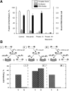
Figure 4
Figure 4
Frozen intermediate of fusion is subsequent to a low pH–triggered change in HA conformation (A) and the LPC-arrested stage (B). (A) FIF is subsequent to the change in fusion sensitivity to neuraminidase and proteinase K upon low pH refolding of HA. Fusion of HAb2 cells with bound RBC was triggered by a 10-min pH 4.9 pulse applied at 4°C. After 45 min at neutral pH at 4°C, cells were warmed to 37°C. During the incubation at neutral pH, 4°C, cells were treated by proteinase K (0.1 mg/ml, 45 min), neuraminidase (0.2 mg/ml, 45 min), or both enzymes simultaneously. In controls, no enzymes were applied. Complete fusion (cell pairs with both PKH26 and CF transferred from RBC to HAb2 cells) and hemifusion (cell pairs with PKH26 but not CF transferred) were assayed 10 min after warming to 37°C using fluorescence microscopy. The binding of RBC to HAb2 cells (righthand y-axis) is presented as the ratio of the number of bound RBC to the number of HAb2 cells at 4°C. Each bar is mean ± SE, n > 3. In the control experiments, we verified that incubation of cells with proteinase K (0.1 mg/ml, 45 min, 4°C) before low-pH application affects neither binding nor complete fusion and unrestricted hemifusion (see also Chernomordik et al., 1997). (B) FIF is subsequent to LPC-arrested stage. Fusion of HAb2 cells (depicted as large ellipses in the illustration of the experimental protocol and result) with bound R18-labeled RBC (two small, shaded ellipses) was triggered by a 10-min pulse of pH 5.0 medium, followed by reneutralization. Final fusion extent was assayed at neutral pH by spectrofluorometry as R18 dequenching. (I) Fusion of cells released from LPC-S was arrested at 4°C. Cells were treated by low pH at 22°C in the presence of 140 μM lauroyl LPC to establish the LPC-arrested stage. 10 min after the end of the pH pulse, cells were placed into a cuvette containing LPC-free solution at either 4 or 22°C. (II) LPC did not block fusion of cells released from FIF. At the end of the pH pulse at 4°C, LPC was added to cells to 180 μM final concentration, still at 4°C. 10 min later, cells were transferred into a cuvette, and fusion was assayed at 22°C in the presence or absence of LPC. We used higher concentrations of LPC at 4°C than at 22°C to achieve the same incorporation of LPC. (III) LPC block prevented the system from reaching FIF. Fusion was triggered at 4°C in the presence of 180 μM lauroyl LPC. 10 min after the end of the low-pH pulse, cells were transferred into a cuvette, and fusion was assayed at 22°C either in the presence or absence of LPC.
Figure 3
Figure 3
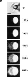
Frozen intermediate of fusion precedes both lipid mixing and fusion pore opening. (A) Complete fusion and unrestricted hemifusion are both reversibly arrested at 4°C. HA-expressing HAb2 cells or GPI-HA–expressing BHA-PI cells with bound PKH26- and CF-labeled RBC were triggered to fuse at pH 4.9, 10 min, 4°C and then incubated in neutral medium for an additional hour at 4°C. Then, still at neutral pH, the temperature was raised to 37°C. Complete fusion (cell pairs with both PKH26 and CF transferred from RBC to HAb2 cells) and hemifusion (cell pairs with PKH26 but not CF transferred) were assayed just before warming cells, still at 4°C, or 10 min after warming. In controls, fusion was triggered at pH 4.9, 10 min, 37°C. The extent of fusion was measured at neutral pH 10 min after the end of low-pH application. Bars are mean ± SE, n > 3. (B) FIF precedes fusion pore formation. No detectable changes in cell capacitance and fusion pore conductance and thus no fusion pore opening was observed when low pH was applied to a HAb2 cell with two bound RBC at 4°C. Arrow at pH pulse. (C) Bright field and fluorescence microscopy images at different stages of the same experiment. Bright field image was taken before acid medium application. The patch pipette is seen on the right; two RBC stained with both PKH26 and CF are seen attached to the left side of the HAb2 cell. Videoimages of PKH26 and CF fluorescence of these cells were taken at 4°C at the onset of the pH pulse and then 10 min after the end of the electrical recording shown in B, when cells were warmed up to 33°C (e). Bar, 10 μm.
Figure 3
Figure 3
Frozen intermediate of fusion precedes both lipid mixing and fusion pore opening. (A) Complete fusion and unrestricted hemifusion are both reversibly arrested at 4°C. HA-expressing HAb2 cells or GPI-HA–expressing BHA-PI cells with bound PKH26- and CF-labeled RBC were triggered to fuse at pH 4.9, 10 min, 4°C and then incubated in neutral medium for an additional hour at 4°C. Then, still at neutral pH, the temperature was raised to 37°C. Complete fusion (cell pairs with both PKH26 and CF transferred from RBC to HAb2 cells) and hemifusion (cell pairs with PKH26 but not CF transferred) were assayed just before warming cells, still at 4°C, or 10 min after warming. In controls, fusion was triggered at pH 4.9, 10 min, 37°C. The extent of fusion was measured at neutral pH 10 min after the end of low-pH application. Bars are mean ± SE, n > 3. (B) FIF precedes fusion pore formation. No detectable changes in cell capacitance and fusion pore conductance and thus no fusion pore opening was observed when low pH was applied to a HAb2 cell with two bound RBC at 4°C. Arrow at pH pulse. (C) Bright field and fluorescence microscopy images at different stages of the same experiment. Bright field image was taken before acid medium application. The patch pipette is seen on the right; two RBC stained with both PKH26 and CF are seen attached to the left side of the HAb2 cell. Videoimages of PKH26 and CF fluorescence of these cells were taken at 4°C at the onset of the pH pulse and then 10 min after the end of the electrical recording shown in B, when cells were warmed up to 33°C (e). Bar, 10 μm.
Figure 1
Figure 1
HA causes unrestricted hemifusion at suboptimal pH. (A) Fusion of HAb2 cells with bound RBC was triggered by a 10-min application of pH 4.9 medium at 37°C. The extent of fusion was assayed by fluorescence microscopy at neutral pH 20 min after the end of low-pH application as lipid dye, PKH26 (circles), and aqueous dye, CF, (squares) redistribution. (B) Pore conductance and PKH26 flux (fluorescence) in a HAb2/ RBC pair treated by pH 4.9 at 33°C. Arrow indicates start of pH pulse. At these “optimal” conditions, pores opened before transfer of membrane dye. (C and D) Unrestricted hemifusion of HAb2 and RBC at pH 5.3. (C) Increase in the integrated PKH26 fluorescence of the HAb2 membrane is not accompanied by any appearance of fusion pore conductance. Arrow indicates start of pH pulse. (D) Bright field (bf) and fluorescent images for the same HAb2/RBC as in (C) pair at different moments of time, 0 time at start of pH pulse. Patch pipette is seen on the right; a PKH26-labeled RBC is seen on left. This lipid dye is seen to redistribute, although no fusion pores are detected (C). Bar, 10 μm.
Figure 1
Figure 1
HA causes unrestricted hemifusion at suboptimal pH. (A) Fusion of HAb2 cells with bound RBC was triggered by a 10-min application of pH 4.9 medium at 37°C. The extent of fusion was assayed by fluorescence microscopy at neutral pH 20 min after the end of low-pH application as lipid dye, PKH26 (circles), and aqueous dye, CF, (squares) redistribution. (B) Pore conductance and PKH26 flux (fluorescence) in a HAb2/ RBC pair treated by pH 4.9 at 33°C. Arrow indicates start of pH pulse. At these “optimal” conditions, pores opened before transfer of membrane dye. (C and D) Unrestricted hemifusion of HAb2 and RBC at pH 5.3. (C) Increase in the integrated PKH26 fluorescence of the HAb2 membrane is not accompanied by any appearance of fusion pore conductance. Arrow indicates start of pH pulse. (D) Bright field (bf) and fluorescent images for the same HAb2/RBC as in (C) pair at different moments of time, 0 time at start of pH pulse. Patch pipette is seen on the right; a PKH26-labeled RBC is seen on left. This lipid dye is seen to redistribute, although no fusion pores are detected (C). Bar, 10 μm.
Figure 2
Figure 2
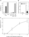
Decreasing either the number of trypsin-activated HA or the temperature results in unrestricted hemifusion. (A) The surface density of fusion-competent HA molecules was varied by altering the duration of trypsin treatment (1 μg/ml, room temperature) of HAb2 cells, which cleaves inactive HA0 into HA1-S-S-HA2 form. Fusion of HAb2 cells with bound RBC was triggered by pH 4.9 medium (10 min, 37°C). Extent of fusion was assayed by fluorescence microscopy at neutral pH 20 min after the end of low-pH application as PKH26 (circles) and CF (squares) redistribution. (B and C) Unrestricted hemifusion of RBC to HAb2 cells mildly treated by trypsin (1 μg/ml, 2 min, room temperature) was triggered by a pH 4.9 pulse applied at 33°C. (B) The fusion pore conductance and the integrated fluorescence of HAb2 membrane for the cell pair in C, arrow at pH pulse. (C) The same cell pair as in B, bright field (bf) and fluorescent images of the HAb2/RBC cell pair at the times indicated. 0 time was at the beginning of the pH pulse. Patch pipette is seen on the right, overlying a PKH26-labeled RBC. This lipid dye is seen to redistribute, although no fusion pores were detected (see B). (D) Nonexpanding pores. When HA is triggered by suboptimal conditions, pores can form that are either transient or fail to expand. Here we triggered with pH 4.9 at 22°C. The pore opened (arrow) 217 s after low-pH application. Nonexpanding pores like this were seen in all conditions (Table I). (E) Unrestricted hemifusion is not an intermediate to complete fusion. Fusion of HAb2 cells with PKH26- and CF-labeled RBC membranes was triggered by a 10-min application of pH 4.9 medium at 37 or 20°C. After low-pH application at 20°C, cells were kept in neutral medium for 10 min more at 20 or 37°C, and then fusion extents were assayed by fluorescence microscopy as redistribution of PKH26 and CF (open and closed bars, respectively). Bar, 10 μm.
Figure 2
Figure 2
Decreasing either the number of trypsin-activated HA or the temperature results in unrestricted hemifusion. (A) The surface density of fusion-competent HA molecules was varied by altering the duration of trypsin treatment (1 μg/ml, room temperature) of HAb2 cells, which cleaves inactive HA0 into HA1-S-S-HA2 form. Fusion of HAb2 cells with bound RBC was triggered by pH 4.9 medium (10 min, 37°C). Extent of fusion was assayed by fluorescence microscopy at neutral pH 20 min after the end of low-pH application as PKH26 (circles) and CF (squares) redistribution. (B and C) Unrestricted hemifusion of RBC to HAb2 cells mildly treated by trypsin (1 μg/ml, 2 min, room temperature) was triggered by a pH 4.9 pulse applied at 33°C. (B) The fusion pore conductance and the integrated fluorescence of HAb2 membrane for the cell pair in C, arrow at pH pulse. (C) The same cell pair as in B, bright field (bf) and fluorescent images of the HAb2/RBC cell pair at the times indicated. 0 time was at the beginning of the pH pulse. Patch pipette is seen on the right, overlying a PKH26-labeled RBC. This lipid dye is seen to redistribute, although no fusion pores were detected (see B). (D) Nonexpanding pores. When HA is triggered by suboptimal conditions, pores can form that are either transient or fail to expand. Here we triggered with pH 4.9 at 22°C. The pore opened (arrow) 217 s after low-pH application. Nonexpanding pores like this were seen in all conditions (Table I). (E) Unrestricted hemifusion is not an intermediate to complete fusion. Fusion of HAb2 cells with PKH26- and CF-labeled RBC membranes was triggered by a 10-min application of pH 4.9 medium at 37 or 20°C. After low-pH application at 20°C, cells were kept in neutral medium for 10 min more at 20 or 37°C, and then fusion extents were assayed by fluorescence microscopy as redistribution of PKH26 and CF (open and closed bars, respectively). Bar, 10 μm.
Figure 5
Figure 5
The stage arrested at 4°C is restricted hemifusion between fusing membranes. (A) Application of hypotonic osmotic shock at FIF but not at LPC-S results in lipid mixing. HAb2 cells with bound R18-labeled RBC were triggered to fuse by pH 5.0, 10 min at 4°C (to establish FIF) or at 22°C with 140 μM lauroyl LPC (to establish LPC-S). Final fusion extent was assayed at neutral pH by spectrofluorometry at 4°C for FIF or with LPC for LPC-S. Application of 4°C, low-tonicity medium to FIF resulted in significant R18 dequenching (more than 60% of that observed in PBS upon warming cells to 22°C). In contrast, almost no R18 dequenching was observed when low-tonicity medium containing 140 μM lauroyl LPC was applied to cells at the LPC-arrested stage (less than 6% of that observed upon washing out LPC by PBS). (B) Chlorpromazine treatment transforms FIF into complete fusion. HAb2 cells with bound, PKH26- and CF-labeled RBC were triggered to fuse at pH 4.9, 10 min, 4°C, incubated at neutral pH, 4°C for 5 min, and then exposed to 0.5 mM CPZ for 2 min, neutral pH, still at 4°C. Complete fusion (cell pairs with both PKH26 and CF transferred from RBC to HAb2 cells) and hemifusion (cell pairs with PKH26 but not CF transferred) were assayed with fluorescence microscopy, still at 4°C. In controls, where cells were treated by pH 4.9, 10 min, 4°C, incubated at neutral pH, 4°C for 10 min, and then warmed up to 37°C, fusion and hemifusion extents assayed 10 min after warming to 37°C were 81 and 17%, respectively. Each bar is mean ± SE, n > 3. (C) Lipid mixing at FIF is promoted by mild treatment with proteinase K. HAb2 cells with bound, doubly labeled RBC were treated at pH 4.9, 10 min, 4°C, incubated for 20 min at neutral pH at 4°C, and then treated with proteinase K (0.05 mg/ml; 4°C) for different times. 1 h after the end of low-pH application, fusion was assayed with fluorescence microscopy, still at 4°C. While no transfer of CF was detected for any of these conditions, proteinase K treatment resulted in a significant percentage of hemifusion events (cell pairs with PKH26 transferred). Each point is mean ± SE, n > 3. Warming the cells at this point yields unrestricted hemifusion (not shown, see Fig. 6 C). Because the proteinase K application (0.05 mg/ml, 15 min, 4°C or 0.1 mg/ml, 45 min, 4°C) to HAb2 cells with bound RBC before low-pH pulse (pH 4.9, 10 min, 4°C) did not result in any measurable transfer of membrane or aqueous dyes at FIF (not shown), the facilitation of the lipid flow at FIF by mild proteinase K treatment is caused by cleaving HA in its low-pH conformation rather than by some nonspecific effects of this enzymatic treatment.
Figure 6
Figure 6
Redirecting cells committed to unrestricted hemifusion toward complete fusion and vice versa. (A and B) HAb2 cells were redirected from unrestricted hemifusion to complete fusion by preventing premature fusion with LPC (A) or with 4°C (B). Each bar is the mean ± SE, n > 3. (A) The percentage of cells committed to complete fusion versus unrestricted hemifusion increased if the onset of actual membrane merger was delayed by applying an LPC block. Fusion was triggered by pH 4.9, 1 min, 22°C without LPC or with 170 μM lauroyl LPC. After a 10-min incubation at neutral pH, LPC was removed by washing cells with LPC-free PBS. Fusion was assayed at neutral pH 20 min after the end of low-pH application. (B) The percentage of cells committed to complete fusion increased with time at 4°C. Fusion was triggered by pH 5.3 medium, 10 min, 4°C. Cells were incubated at neutral pH at 4°C for different time intervals and finally warmed up to 37°C, still at neutral pH. Fusion was assayed 10 min after warming to 37°C. In controls, fusion was triggered by pH 5.3 medium, 10 min, 37°C. Fusion extents were measured at neutral pH 20 min after the end of low-pH application. (C) Cells committed to complete fusion but blocked at FIF could still be redirected to unrestricted hemifusion by cleaving some activated HA molecules. Fusion was triggered by pH 4.9 medium, 10 min, 4°C. Cells were incubated in neutral pH medium still at 4°C, 30 min and then warmed to 37°C. 20 min later fusion was assayed. In the time interval between the end of the low-pH application and warming, cells were treated with proteinase K (0.1 mg/ml, 4°C) for different times. Complete fusion (cell pairs with both PKH26 and CF transferred from RBC to HAb2 cells) and hemifusion (cell pairs with PKH26 but not CF transferred) were assayed with fluorescence microscopy. Each point is mean ± SE, n > 3.
Figure 7
Figure 7
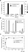
Promotion and inhibition of complete fusion by altering membrane lipid composition. (A) LPC in the inner monolayer of RBC membrane promotes complete fusion. Fusion was triggered at 22°C, pH 5.1, 10 min. No exogenous lipid was added in the control experiments. Lauroyl LPC was either added to the medium to incorporate into outer membrane monolayers (170 μM), loaded into RBC to partition into inner membrane monolayer, or both loaded into RBC and added to the outer membrane monolayers (170 μM). (B) Loading LPC into RBC ghosts decreases the number of activated HA molecules required for fusion pore formation and expansion, downstream of FIF. Fusion of HAb2 cells with either RBC loaded with LPC or control RBC was triggered at 4°C, pH 4.9, 10 min. Then cells were incubated in neutral pH medium for 10 min, treated by proteinase K (0.1 mg/ ml, still at 4°C) for 45 s, 150 s, or not treated at all. After 20 min more at neutral pH, 4°C, the temperature was raised to 22°C. (C) Complete fusion is inhibited by OA. Fusion was triggered at 22°C, pH 4.9, 10 min in the medium containing different concentrations of OA. In A–C, complete fusion (cell pairs with both PKH26 and CF transferred from RBC to HAb2 cells) and hemifusion (cell pairs with PKH26 but not CF transferred) were assayed with fluorescence microscopy after 20 min of incubation at neutral pH. Each point is mean ± SE, n > 3.
Figure 8
Figure 8
Pathway for the fusion of HA- expressing membrane with RBC membrane. In the schematic diagram of the fusion site, the top membrane is a section of an HA-expressing cell, transmembrane domain of HA pointing up. The bottom membrane is a section of a target cell membrane (e.g., RBC), fusion peptide pointing down. A1, B1, C1, D, and E illustrate the pathway of complete fusion. Acidification causes a conformational change in HA from its initial state (A1), leading to insertion of the HA fusion peptide into the target membrane. (B1) The fusion intermediate after assembly of refolded HA but before membrane merger, which is the LPC-arrested state (LPC-S). (C1) Restricted hemifusion intermediate within a fence of activated HA, which restricts lipid diffusion between the two membranes: the stage arrested at 4°C (FIF). (D) Formation of a fusion pore, which can close and open again within the fence (flicker). (E) Expansion of the fusion pore beyond the fence to complete fusion. The lower set of intermediates (A2–F) illustrates the unrestricted hemifusion pathway for decreased surface density of activated HA near the fusion site. (A2) Initial state. (B2) LPC-arrested state. (C2) Restricted hemifusion intermediate arrested at 4°C (FIF). Note that the fence of activated HA assembled the around fusion site, which is formed here by a lower number of proteins than the one in C1, still restricts lipid transfer between the two membranes. (F) The extension of the hemifusion intermediate precedes the assembly of a proper HA complex and yields an unrestricted hemifusion state that allows dye mixing but is incapable of supporting fusion pore development. Blocking the premature merger of membranes and development of unrestricted hemifusion with either LPC (at B2) or 4°C (at C2) allows activated HA molecules to assemble over a longer period of time and thus redirects the cells from the unrestricted hemifusion pathway to the complete fusion pathway. In contrast, cleaving of some of the activated HA molecules at C1 with proteinase K redirects cells from the complete fusion pathway to the unrestricted hemifusion pathway.

References

    1. Anderson CM, Georgiou GN, Morrison IE, Stevenson GV, Cherry RJ. Tracking of cell surface receptors by fluorescence digital imaging microscopy using a charge-coupled device camera. Low-density lipoprotein and influenza virus receptor mobility at 4°C. J Cell Sci. 1992;101:415–425. - PubMed
    1. Bagai S, Lamb RA. Truncation of the COOH-terminal region of the paramyxovirus SV5 fusion protein leads to hemifusion but not complete fusion. J Cell Biol. 1996;135:73–84. - PMC - PubMed
    1. Bentz J, Ellens H, Alford D. An architecture for the fusion site of influenza hemagglutinin. FEBS Lett. 1990;276:1–5. - PubMed
    1. Bierbaum TJ, Bouma SR, Huestis WH. A mechanism of erythrocyte lysis by lysophosphatidylcholine. Biochim Biophys Acta. 1979;555:102–110. - PubMed
    1. Blumenthal R, Pak CC, Raviv Y, Krumbiegel M, Bergelson LD, Morris SJ, Lowy RJ. Transient domains induced by influenza haemagglutinin during membrane fusion. Mol Membr Biol. 1995;12:135–142. - PubMed

MeSH terms

Substances