. 1998 Mar 23;140(6):1407-16.
doi: 10.1083/jcb.140.6.1407.

Role of the kinesin neck region in processive microtubule-based motility

Affiliations

Role of the kinesin neck region in processive microtubule-based motility

L Romberg et al. J Cell Biol. .

Abstract

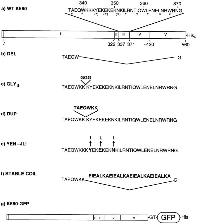
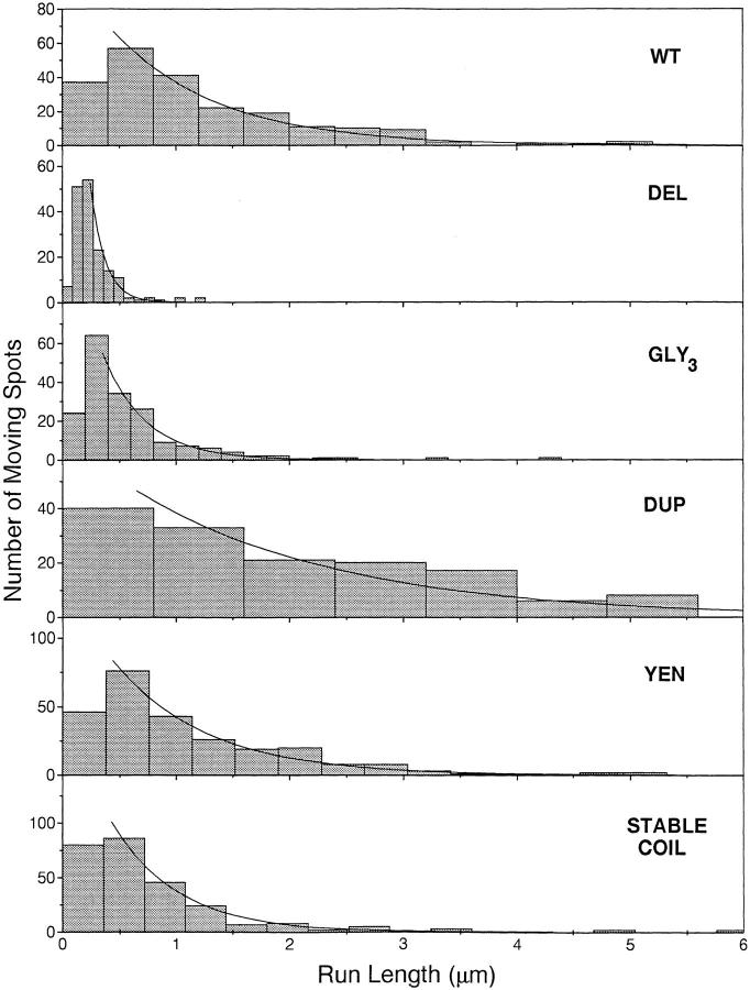
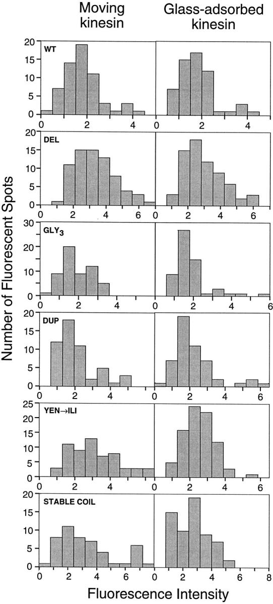
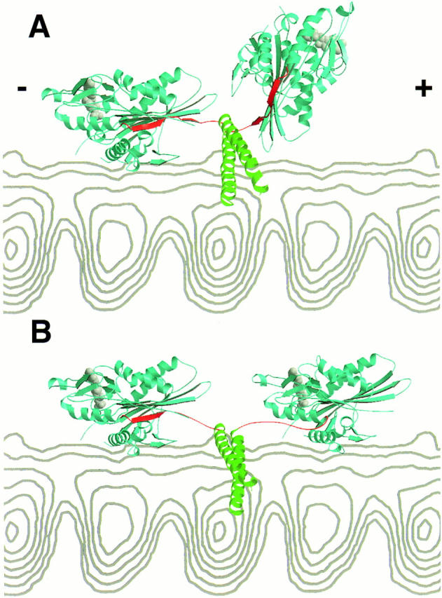
Kinesin is a dimeric motor protein that can move along a microtubule for several microns without releasing (termed processive movement). The two motor domains of the dimer are thought to move in a coordinated, hand-over-hand manner. A region adjacent to kinesin's motor catalytic domain (the neck) contains a coiled coil that is sufficient for motor dimerization and has been proposed to play an essential role in processive movement. Recent models have suggested that the neck enables head-to-head communication by creating a stiff connection between the two motor domains, but also may unwind during the mechanochemical cycle to allow movement to new tubulin binding sites. To test these ideas, we mutated the neck coiled coil in a 560-amino acid (aa) dimeric kinesin construct fused to green fluorescent protein (GFP), and then assayed processivity using a fluorescence microscope that can visualize single kinesin-GFP molecules moving along a microtubule. Our results show that replacing the kinesin neck coiled coil with a 28-aa residue peptide sequence that forms a highly stable coiled coil does not greatly reduce the processivity of the motor. This result argues against models in which extensive unwinding of the coiled coil is essential for movement. Furthermore, we show that deleting the neck coiled coil decreases processivity 10-fold, but surprisingly does not abolish it. We also demonstrate that processivity is increased by threefold when the neck helix is elongated by seven residues. These results indicate that structural features of the neck coiled coil, although not essential for processivity, can tune the efficiency of single molecule motility.


Figures

Figure 1
Figure 1
Mutant neck constructs. (a) WT K560: The wild-type kinesin construct used as the basis for mutagenesis contains the first 560 amino acids of the human kinesin gene followed by a histidine tag. *, residues at the hydrophobic interface of the coiled coil; (*), destabilizing residues in this interface. Domains of this construct are (I) core catalytic domain, (II) neck β sheet region, (III) neck coiled-coil region, (IV) hinge region, (V) coiled-coil stalk. The boundaries of the core catalytic domain are defined by conservation throughout the kinesin superfamily. The boundary of the neck is defined by strong class-specific conservation among conventional kinesins (Vale and Fletterick, 1997). (b) DEL: aa 341–370 of the neck coiled coil were deleted. (c) GLY3: three glycines were inserted between K342 and K343. (d) DUP: Residues T336–K342, one complete turn of the α-helix, were duplicated. (e) YEN→ ILI: Three destabilizing residues at the “a” and “d” position of the coiled coil were changed to stabilizing hydrophobic residues (Y344I, E347L, and N351I). (f) STABLE COIL: Four heptad repeats (aa 343–370), were replaced by a highly stable model coiled coil consisting of four repeats of the sequence EIEALKA. (g) WT K560–GFP: The above neck mutations (b–f) were also inserted into K560 with GFP, the Ser65Thr mutant (Heim et al., 1995), fused to its COOH terminus.
Figure 3
Figure 3
Run lengths of single, fluorescently labeled kinesin molecules. Run lengths of 150–270 individual GFP–kinesin molecules moving on axonemes were measured from two independent preparations of each construct. Histograms of the data were plotted using bin widths derived from the formula 2.6σn(−1/3) (Scott, 1979), where σ is the standard deviation of the data and n is the number of data points collected. Exponential curves were fit to the data using only runs >0.25 μm (or 0.2 and 0.15 μm for GLY3 and DEL, respectively), as described in Materials and Methods. Run-length values are shown in Table II.
Figure 2
Figure 2
Fluorescent intensity of individual kinesin molecules. The histograms show the fluorescent intensity of kinesin molecules either moving along axonemes or nonspecifically adsorbed onto the slide surface nearby (refer to Materials and Methods for details). Tick marks represent one arbitrary fluorescent unit; fluorescent intensities cannot be directly compared between preparations because of small variations in laser alignment during different assays.
Figure 4
Figure 4
A structural model for how the kinesin dimer might span the eight nanometers between adjacent α/β tubulin binding sites. In this crystal structure of the rat kinesin dimer (Kozielski et al., 1997), the catalytic core domain is colored blue, the nucleotide is colored gray, the β strand region of the neck (β9 and β10; rat aa 321–336) is colored red, and the neck coiled coil (rat aa 337–370) is colored green (note: the rat kinesin aa numbers differ by −2 aa compared to human kinesin in this region). A side view of a microtubule protofilament from cryoelectron microscopy reconstructions (Hoenger et al., 1995) is shown in gray. The microtubule plus end (the direction of travel for kinesin) is at the right. In A, the unaltered crystal structure of the rat kinesin dimer is shown with one head docked onto the microtubule. The approximate orientation of the bound head was defined by having the half of the molecule containing the nucleotide pointing towards the minus end, the “arrowhead tip” pointing towards the plus end (Hoenger and Milligan, 1997; Sosa et al., 1997), and the main microtubule binding loop (L12) in contact with tubulin surface (Sosa et al., 1997; Woehlke et al., 1997). As noted by Kozielski et al. (1997), the neck coiled coil runs perpendicular to the long axis of the protofilament and is located near but not sterically clashing with the microtubule surface. In the crystal structure, the distance between the two heads is insufficient to enable the second head to dock onto the microtubule. It is important to mention that the structure shown here may not exactly correspond to one that occurs normally in the motility cycle, since the geometry of the heads could be partially determined by crystal contacts and since microtubule or nucleotide binding may change the solution conformation. In B, the β strands between aa 327–336 were separated from the catalytic core in the leading head using the program O (T.A. Jones and M. Kjeldgaard), obeying restraints of bond distances and geometries. This generates a sufficiently long linker to enable the leading head to dock to the adjacent tubulin binding site in the identical orientation to the lagging head. Only modest adjustments need to be made to the neck β strands of the lagging head, since they are already extended and pointing towards the microtubule plus end. In this model, the neck coiled coil does not unwind. The nucleotide (ADP) from the crystal structure is shown in both heads in these panels, although it is more likely that the two heads are in different nucleotide states during the motility cycle.

Similar articles

Cited by

References

    1. Amos LA, Hirose K. The structure of microtubule-motor complexes. Curr Opin Cell Biol. 1997;9:4–11. - PubMed
    1. Arnal I, Metoz F, DeBonis S, Wade RH. Three-dimensional structure of functional motor proteins on microtubules. Curr Biol. 1996;6:1265–1270. - PubMed
    1. Berliner E, Young EC, Anderson K, Mahtani H, Gelles J. Failure of a single-headed kinesin to track parallel to microtubule protofilaments. Nature. 1995;373:718–721. - PubMed
    1. Block SM. Fifty ways to love your lever: myosin motors. Cell. 1996;87:151–157. - PubMed
    1. Block SM, Goldstein LS, Schnapp BJ. Bead movement by single kinesin molecules with optical tweezers. Nature. 1990;348:348–352. - PubMed

Publication types