. 1998 Mar 23;140(6):1427-39.
doi: 10.1083/jcb.140.6.1427.

A requirement for cyclin D3-cyclin-dependent kinase (cdk)-4 assembly in the cyclic adenosine monophosphate-dependent proliferation of thyrocytes

Affiliations

A requirement for cyclin D3-cyclin-dependent kinase (cdk)-4 assembly in the cyclic adenosine monophosphate-dependent proliferation of thyrocytes

F Depoortere et al. J Cell Biol. .

Abstract

In different systems, cyclic adenosine monophosphate (cAMP) either blocks or promotes cell cycle progression in mid to late G1 phase. Dog thyroid epithelial cells in primary culture constitute a model of positive control of DNA synthesis initiation and G0-S prereplicative phase progression by cAMP as a second messenger for thyrotropin (TSH). The cAMP-dependent mitogenic pathway is unique as it is independent of mitogen-activated protein kinase activation and differs from growth factor-dependent pathways at the level of the expression of several protooncogenes/transcription factors. This study examined the involvement of D-type G1 cyclins and their associated cyclin-dependent kinase (cdk4) in the cAMP-dependent G1 phase progression of dog thyroid cells. Unlike epidermal growth factor (EGF)+serum and other cAMP-independent mitogens, TSH did not induce the accumulation of cyclins D1 and D2 and partially inhibited the basal expression of the most abundant cyclin D3. However, TSH stimulation enhanced the nuclear detection of cyclin D3. This effect correlated with G1 and S phase progression. It was found to reflect both the unmasking of an epitope of cyclin D3 close to its domain of interaction with cdk4, and the nuclear translocation of cyclin D3. TSH and EGF+serum also induced a previously undescribed nuclear translocation of cdk4, the assembly of precipitable cyclin D3-cdk4 complexes, and the Rb kinase activity of these complexes. Previously, cdk4 activity was found to be required in the cAMP-dependent mitogenic pathway of dog thyrocytes, as in growth factor pathways. Here, microinjections of a cyclin D3 antibody showed that cyclin D3 is essential in the TSH/ cAMP-dependent mitogenesis, but not in the pathway of growth factors that induce cyclins D1 and D2. The present study (a) provides the first example in a normal cell of a stimulation of G1 phase progression occurring independently of an enhanced accumulation of cyclins D, (b) identifies the activation of cyclin D3 and cdk4 through their enhanced assembly and/or nuclear translocation, as first convergence steps of the parallel cAMP-dependent and growth factor mitogenic pathways, and (c) strongly suggests that this new mechanism is essential in the cAMP-dependent mitogenesis, which provides the first direct demonstration of the requirement for cyclin D3 in a G1 phase progression.


Figures
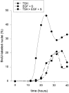
Figure 1
Figure 1
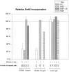
Kinetics of cell cycle progression after mitogenic stimulation of quiescent dog thyrocytes. 4-d-old dog thyrocytes were stimulated at time 0 by TSH (1 mU/ml), EGF (25 ng/ml)+serum (10%), or a combination of TSH, EGF, and serum (10%). 1 h before fixation at the times indicated, cells were incubated with BrdU. BrdU incorporation was detected by immunofluorescent staining and the percentage of cells in S phase was determined.
Figure 2
Figure 2
Western blotting analyses of the accumulation of the three cyclins D in dog thyrocytes after stimulation by different mitogenic treatments. Cells remained quiescent in control conditions (C) or were stimulated by TSH (1 mU/ml), EGF (25 ng/ ml)+serum (10%; ES), or the combination of these factors (ES+TSH) for 8 to 32 h. (A) Immunoblot autoradiograph. 30 μg of cellular proteins were loaded per lane. The exposure durations were different for the autoradiographical detection of the different cyclins D: cyclin D1 (DCS-6), 8 d; cyclin D2 (DCS-3), 16 d; cyclin D3 (DCS-22), 2 d. ND, not done or lost. (B) Detection of cyclin D3 with DCS-29 from control cells (C) and cells stimulated by TSH (T) or ES for 20 h. This autoradiograph was exposed for 8 d like the cyclin D1 immunoblot shown in A, in order to illustrate the relative abundances of cyclin D3 and cyclin D1. It also shows the high specificity of the DCS-29 antibody used in the microinjection experiments of Figs. 10 and 11. (C) Densitometry quantitation of cyclin D1 from the autoradiograph details in A. (D) Densitometry quantitation of cyclin D3 (shown with a more precise kinetics from another dog thyrocyte primary culture in order to illustrate the qualitative reproducibility compared with the results in A). In C and D, the relative amount of cyclin D1 or cyclin D3 after cell stimulation is expressed compared with the average (fixed as 100) of values obtained at different times from unstimulated (control) cells in the same experiments.
Figure 2
Figure 2
Western blotting analyses of the accumulation of the three cyclins D in dog thyrocytes after stimulation by different mitogenic treatments. Cells remained quiescent in control conditions (C) or were stimulated by TSH (1 mU/ml), EGF (25 ng/ ml)+serum (10%; ES), or the combination of these factors (ES+TSH) for 8 to 32 h. (A) Immunoblot autoradiograph. 30 μg of cellular proteins were loaded per lane. The exposure durations were different for the autoradiographical detection of the different cyclins D: cyclin D1 (DCS-6), 8 d; cyclin D2 (DCS-3), 16 d; cyclin D3 (DCS-22), 2 d. ND, not done or lost. (B) Detection of cyclin D3 with DCS-29 from control cells (C) and cells stimulated by TSH (T) or ES for 20 h. This autoradiograph was exposed for 8 d like the cyclin D1 immunoblot shown in A, in order to illustrate the relative abundances of cyclin D3 and cyclin D1. It also shows the high specificity of the DCS-29 antibody used in the microinjection experiments of Figs. 10 and 11. (C) Densitometry quantitation of cyclin D1 from the autoradiograph details in A. (D) Densitometry quantitation of cyclin D3 (shown with a more precise kinetics from another dog thyrocyte primary culture in order to illustrate the qualitative reproducibility compared with the results in A). In C and D, the relative amount of cyclin D1 or cyclin D3 after cell stimulation is expressed compared with the average (fixed as 100) of values obtained at different times from unstimulated (control) cells in the same experiments.
Figure 10
Figure 10
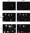
Requirement of cyclin D3 for G1 phase progression stimulated by TSH, but not by the cAMP-independent mitogen HGF. Quiescent dog thyrocytes were injected with the DCS-29 cyclin D3 monoclonal antibody (6 mg/ml), or with the DCS-6 cyclin D1 blocking antibody, and stimulated for 40 h with TSH (1 mU/ml) or HGF (50 ng/ml). BrdU was added 24 h before fixation. Nuclei were identified by Hoechst 33342 staining of DNA. Microinjected cells were identified by the immunodetection of the injected antibody (IgG). BrdU incorporation was coimmunodetected (BrdU). Arrowheads indicate injected cells that have incorporated BrdU. Note that none of the TSH-stimulated cells injected with the DCS-29 cyclin D3 antibody incorporated BrdU, in contrast to neighboring noninjected cells, TSH-stimulated cells injected with the DCS-6 cyclin D1 antibody, and HGF-stimulated cells injected with the DCS-29 cyclin D3 antibody.
Figure 11
Figure 11
Summary of cyclin D3 antibody microinjection experiments. Quiescent dog thyrocytes were injected by 2 or 6 mg/ml DCS-29 cyclin D3 antibody or a control IgG and processed for immunofluorescent detection of injected immunoglobulin and BrdU as in Fig. 10, after stimulation for 40 h as indicated, with TSH (1 mU/ml), forskolin (Fo; 10−5M), HGF (50 ng/ml), EGF (25 ng/ml)+serum (S), or EGF alone. Results are expressed (mean+SD) relative to the percentages of BrdU-labeled nuclei in neighboring noninjected cells. Average percentage of BrdU-labeled nuclei in noninjected cells were 37% (TSH), 50% (Fo), 46% (HGF), 69% (EGF+S), 35% (EGF), and 3% in control unstimulated cells. The number of separate experiments and the total number of injected cells analyzed are indicated for each condition. In each separate experiment, the stimulation by TSH (or Fo) was compared with one of the cAMP-independent stimulations (HGF, EGF+S, or EGF).
Figure 3
Figure 3
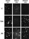
Immunofluorescence labeling of cyclin D1 (DCS-6), cyclin D2 (DCS-3), and cyclin D3 (DCS-22) in dog thyrocytes stimulated by different mitogenic treatments. Quiescent 4-d-old cells were stimulated for 20 h by TSH (1 mU/ml), EGF (25 ng/ ml)+serum (10%; ES), or remained in control condition (C). Exposure times of the photographs were the same for the different cell treatments, but were shorter for cyclin D3 than cyclin D1 and much longer for cyclin D2.
Figure 4
Figure 4
Correlation between the effects of various mitogenic treatments on DNA synthesis and on the increase of cyclin D3 reactivity to DCS-22. 4-d-old quiescent dog thyrocytes were stimulated with either TSH (1 mU/ml), EGF (25 ng/ml; E), TPA (10 ng/ml; TPA), HGF (40 ng/ml; H), EGF+serum (10%; ES) or remained in the usual control conditions in the presence of insulin (5 μg/ml; C). Some cells were maintained since the beginning of the culture in the absence of insulin (α). 20 h after stimulation (i.e., when a maximum of cells are in mid to late G1), cells were fixed and processed for cyclin D3 immunofluorescent labeling using DCS-22. The nuclear fluorescence was measured over 200 cells per conditions. For each condition, the proliferative activity was evaluated 48 h after mitogenic stimulation by the incorporation of BrdU during the last 24 h of incubation, and plotted as a function of the average intensity of cyclin D3 immunofluorescence.
Figure 5
Figure 5
Double immunofluorescence labeling of cyclin D3 (detected using DCS-22), and PCNA used as a cell cycle marker in the same cells. Before fixation, 4-d-old dog thyrocytes were stimulated for 26 h with TSH (1 mU/ml), EGF (25 ng/ml)+serum (10%; ES), or remained quiescent in control medium (C) for this time. Arrowheads and large arrows show cells identified, respectively, in late G1 and S phase as described previously (Baptist et al., 1993, 1996). Small arrows show G0/G1 cells with a low PCNA staining.
Figure 6
Figure 6
Immunofluorescence labeling of cyclin D3 in dog thyrocytes. Quiescent 4-d-old cells were stimulated for 20 h by TSH (1 mU/ml) or remained in control condition (C). Cyclin D3 was detected in the same experiment using DCS-22 or DCS-28 as in Fig. 3, or using DCS-22 after a retrieval treatment of masked antigen by trypsin. All the DCS-22 labelings were photographed with the same exposure time. Note the more intense staining of control cells with DCS-28 or with DCS-22 after the trypsin preincubation, and in these two last conditions, the increase of the nuclear staining of cyclin D3 at the expense of its cytoplasmic staining in many TSH-stimulated cells, which suggests a nuclear translocation.
Figure 7
Figure 7
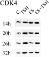
Time course of the accumulation of cdk4 in dog thyrocytes after stimulation by different mitogenic treatments. Cells were stimulated by TSH (1 mU/ ml), EGF (25 ng/ml)+serum (10%; ES), or the combination of these factors (ES+TSH) for 14–32 h. Some cells remained quiescent in control conditions (C) and were analyzed at 14 and 26 h. Autoradiography of cdk4 detection by Western blotting. ND, not done.
Figure 8
Figure 8
Immunofluorescence labeling of cdk4 in dog thyrocytes. Before fixation, 4-d-old dog thyrocytes were stimulated for 20 h with TSH (1 mU/ml), EGF (25 ng/ml)+serum (10%; ES), or remained quiescent in control condition (C). Note the increase of nuclear staining of cdk4 at the expense of its cytoplasmic staining in many stimulated cells, which suggests a nuclear translocation.
Figure 9
Figure 9
Convergence of cAMP-dependent and -independent mitogenic treatments on cyclin D3–cdk4 complex formation and Rb phosphorylation. 4-d-old dog thyrocytes were stimulated for 20 h with either TSH (1 mU/ml; T), EGF (25 ng/ml)+serum (10%; ES), or remained quiescent in control medium (C) for this time. (A) Assembly of cyclin D3–cdk4 complexes. The complexes were immunoprecipitated as indicated from equal amounts of cell extract using either DCS-28 (cyclin D3) or DCS-35 (cdk4) monoclonal antibodies, and the presence of cyclin D3 and cdk4 was detected by Western blot in these immunoprecipitates. A same autoradiograph exposure is provided throughout in order to allow the direct comparison of amounts of total versus complexed cdk4 and cyclin D3. (B) Cyclin D3–associated Rb kinase activity using GST-Rb COOH-terminal protein (GST-Rbc) and γ-[32P]ATP as substrates. (C) Phosphorylation of Rb detected by its electrophoretic shift in Western blot of whole cell lysates.

Similar articles

Cited by

References

    1. Ajchenbaum F, Ando K, De Caprio JA, Griffin JD. Independent regulation of human D-type cyclin gene expression during G1 phase in primary human T lymphocytes. J. Biol Chem. 1993;268:4113–4119. - PubMed
    1. Baldin V, Lukas J, Marcotte MJ, Pagano M, Draetta G. Cyclin D1 is a nuclear protein required for cell cycle progression in G1. Genes Dev. 1993;7:812–821. - PubMed
    1. Baptist M, Dumont JE, Roger PP. Demonstration of cell cycle kinetics in thyroid primary culture by immunostaining of proliferating cell nuclear antigen: differences in cyclic AMP-dependent and -independent mitogenic stimulations. J Cell Sci. 1993;105:69–80. - PubMed
    1. Baptist M, Dumont JE, Roger PP. Intercellular heterogeneity of early mitogenic events: cAMP generalizes the EGF effect on c-Fos protein appearance but not on MAP kinase phosphorylation and nuclear translocation in dog thyroid epithelial cells. Exp Cell Res. 1995;221:160–171. - PubMed
    1. Baptist M, Lamy F, Gannon J, Hunt T, Dumont JE, Roger PP. Expression and subcellular localization of CDK2 and cdc2 kinases and their common partner cyclin A in thyroid epithelial cells: comparison of cyclic AMP-dependent and -independent cell cycles. J Cell Physiol. 1996;166:256–273. - PubMed

Publication types

MeSH terms