. 1998 Mar 15;507 ( Pt 3)(Pt 3):759-69.
doi: 10.1111/j.1469-7793.1998.759bs.x.

The effect of tetracaine on stimulated contractions, sarcoplasmic reticulum Ca2+ content and membrane current in isolated rat ventricular myocytes

Affiliations

The effect of tetracaine on stimulated contractions, sarcoplasmic reticulum Ca2+ content and membrane current in isolated rat ventricular myocytes

C L Overend et al. J Physiol. .

Abstract

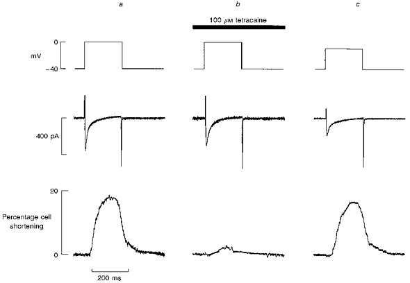
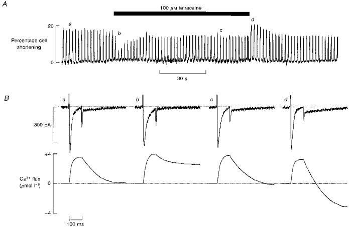
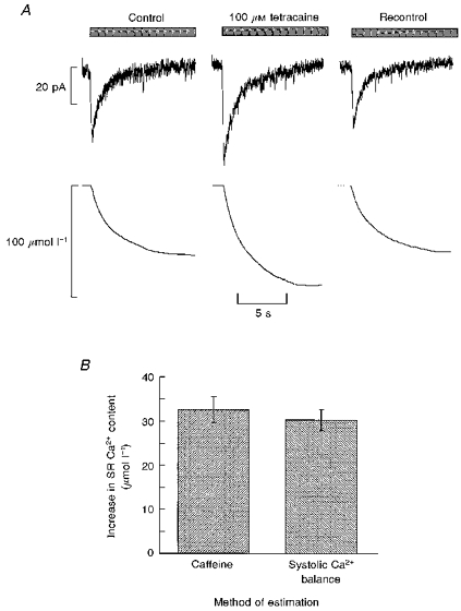
1. The effects of tetracaine were examined on rat ventricular myocytes. In both field-stimulated and voltage-clamped cells tetracaine (100-200 microM) produced an initial decrease of contraction before a recovery towards the control level. Removal of tetracaine produced a transient overshoot of contraction to levels greater than the control. 2. The transient decrease of contraction produced by tetracaine was accompanied by a small transient increase in the integral of the L-type Ca2+ current and a larger transient decrease of the Na+-Ca2+ exchange current on repolarization. These are attributed to decreased systolic release of Ca2+. On removal of tetracaine there was an increase of the Na+-Ca2+ exchange current. Before the addition of tetracaine, calculated Ca2+ influx and efflux across the sarcolemma were approximately equal. On adding tetracaine, efflux was transiently less than influx and, on removal of tetracaine, efflux was greater than influx. 3. These changes in Ca2+ fluxes result in an increase of cell Ca2+ during exposure to tetracaine. The calculated magnitude of this increase was equal to that measured directly by applying caffeine (20 mM) to release sarcoplasmic reticulum (SR) Ca2+ and integrating the resulting Na+-Ca2+ exchange current. 4. It is concluded that the effects of tetracaine can be accounted for by depression of calcium-induced Ca2+ release (CICR). The response is transient because the inhibition is compensated for by an increase of SR Ca2+ content such that there is no steady-state effect on the magnitude of the systolic Ca2+ transient. The consequences of this result for the effects of other modulators of CICR are discussed.


Figures

Figure 1
Figure 1. The effects of tetracaine on contraction amplitude a nd systolic [Ca2+]i
A, time course of cell shortening (top) and indo-1 ratio (R = F400/F500) (bottom). The cell was field stimulated at a frequency of 0.3 Hz. Tetracaine (100 μm) was applied for the period indicated by the bar. The indo-1 ratio was corrected for the intrinsic fluorescence of tetracaine (see Methods). B, averaged specimen (n = 5) Ca2+ transients taken from the periods (a-d) indicated in A.
Figure 2
Figure 2. The effects of tetracaine on contraction and membrane current in a voltage-clamped cell
A, time course of changes of contraction. Tetracaine (100 μm) was applied for the period indicated by the bar. The membrane potential was held at −40 mV, and 200 ms duration depolarizing pulses to 0 mV were applied at a frequency of 0.5 Hz. B, specimen records of membrane current (top) and contraction (bottom) obtained at the times (a-d) indicated in A.
Figure 3
Figure 3. Comparison of the effects of tetracaine with those of decreasing the size of depolarization
In all panels the traces show (from top to bottom): membrane potential, current, cell length. In a and b, the depolarizing pulse was to 0 mV. Panel a, control; b, after 4 s exposure to tetracaine (100 μm); c, in the absence of tetracaine (depolarization to −10 mV). Membrane potential was held at −40 mV and depolarizing pulses of 200 ms duration were applied to elicit contraction.
Figure 4
Figure 4. Transient loss of Ca2+ flux balance during application and removal of tetracaine
A, time course of the effects on contraction of applying tetracaine for the period shown by the bar. B, specimen records of membrane current (top) and cumulative integral (bottom). Membrane potential was held at −40 mV and depolarizing pulses of 100 ms duration were applied at 0.5 Hz. The Ca2+ flux traces show the cumulative integral of the calcium current (initial upward deflection) followed by a downward deflection due to the Ca2+ efflux. The records were obtained at the times (a-d) shown in A. (See Methods for calculation of sacrolemmal Ca2+ movements.) The vertical positions of the current traces have been aligned to facilitate comparison. Tetracaine produced an outward shift of holding current of 7 pA and this has been removed to facilitate comparison between records.
Figure 5
Figure 5. Net accumulation of calcium during exposure to and removal of tetracaine
The traces show (from top to bottom): calculated Ca2+ entry via the L-type Ca2+ current, Ca2+ efflux on repolarization (calculated as shown in Fig. 4), cumulative change of cell Ca2+ content. Calcium content (presumably SR) is expressed per unit total cell volume.
Figure 6
Figure 6. The effect of 200 μm tetracaine on contraction and current
A, time course of cell shortening in response to electrical stimulation elicited by 200 ms depolarizing steps to 0 mV from a holding potential of −40 mV. Tetracaine (200 μm) was applied as indicated by the bar. B, specimen records of membrane current (top) and cumulative integral (bottom). The records were obtained at the times (a-d) shown in A. The records in a and b are the means of 10 and 5 pulses, respectively. Single pulses are shown in c and d.
Figure 7
Figure 7. The gain of measured SR Ca2+ content matches that calculated from the sarcolemmal fluxes
A, measurement of SR Ca2+ content. Caffeine (20 mm) was applied for the period indicated by the bars. Traces show current (top) and integrated current (bottom). From left to right: control; after 1.5 min exposure to tetracaine (100 μm); recontrol (1.5 min after removing tetracaine). B, histogram comparing the measured changes of SR Ca2+ (left) with those calculated as in Fig. 5 from the cumulative integrals (right).

References

    1. Adachi-Akahane S, Cleemann L, Morad M. Cross-signaling between L-type Ca2+ channels and ryanodine receptors in rat ventricular myocytes. Journal of General Physiology. 1996;108:435–454. 10.1085/jgp.108.5.435. - DOI - PMC - PubMed
    1. Almers W, Best PM. Effects of tetracaine on displacement currents and contraction of frog skeletal muscle. Journal of Physiology. 1976;262:583–611. - PMC - PubMed
    1. Bassani JWM, Yuan W, Bers DM. Fractional SR Ca release is regulated by trigger Ca and SR Ca content in cardiac myocytes. American Journal of Physiology. 1995;268:C1313–1319. - PubMed
    1. Bridge JH, Smolley JR, Spitzer KW. The relationship between charge movements associated with ICa and INa-Ca in cardiac myocytes. Science. 1990;248:376–378. - PubMed
    1. Carmeliet E, Morad M, Van der Heyden G, Vereecke J. Electrophysiological effects of tetracaine in single guinea-pig ventricular myocytes. Journal of Physiology. 1986;376:143–161. - PMC - PubMed

MeSH terms

LinkOut - more resources