. 1998 Mar 15;507 ( Pt 3)(Pt 3):783-94.
doi: 10.1111/j.1469-7793.1998.783bs.x.

Glycine-activated currents are changed by coincident membrane depolarization in developing rat auditory brainstem neurones

Affiliations

Glycine-activated currents are changed by coincident membrane depolarization in developing rat auditory brainstem neurones

K H Backus et al. J Physiol. .

Abstract

1. During early ontogeny, glycine receptors (GlyRs) exert depolarizing responses which may be of developmental relevance. We have used the gramicidin-perforated patch technique to elucidate the mechanism of glycine-activated currents in developing neurones of the rat lateral superior olive (LSO). 2. When the holding potential was set to -60 mV, perforated-patch recordings revealed glycine-induced inward currents in 59 %, outward currents in 5 % and biphasic currents in 34 % of the LSO neurones tested (n = 44). The biphasic currents were characterized by a transient outward phase which was followed by an inward phase. 3. Ion substitution experiments showed that both Cl- and HCO3- contributed to the glycine- induced biphasic current responses. 4. In the biphasic responses, the reversal potential of the glycine-induced current (Egly) depended on the response phase. A strong shift of Egly from a mean of -72 mV during the outward phase of the glycine response to a mean of -51 mV during the inward phase was observed, suggesting a shift of an ion gradient. 5. When the membrane potential was depolarized, 'tail' currents were induced in the presence of glycine. An increased duration or amplitude of the evoked depolarizations resulted in a proportional enlargement of these tail currents, indicating that they were produced by a shift of an ion gradient. Since changes of the HCO3- gradient are negligible, because of the carbonic anhydrase activity, we suggest that these tail currents were caused by a shift of the Cl- gradient. 6. We conclude that Cl- accumulates intracellularly during the activation of GlyRs and, consequently, Egly moves towards more positive values. 7. Coincident depolarizing stimuli enhanced intracellular Cl- accumulation and the shift of Egly, thereby switching hyperpolarizing to depolarizing action. This change could assist in an activity-dependent strengthening and refinement of glycinergic synapses during the maturation of inhibitory connectivity.


Figures
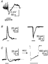
Figure 1
Figure 1. Glycine-activated responses in LSO neurones
Glycine (1 mm) was applied to the bath as indicated by the horizontal bars. A, gramicidin-perforated patch recording in the current-clamp mode showed biphasic membrane potential changes in 50% of neurones. B, when recording in the voltage-clamp mode of the perforated-patch configuration, glycine evoked an outward current in 5% of the neurones (left), but an inward current in 59% (right). C, in 34% of the neurones, glycine induced biphasic current responses in the perforated-patch configuration (left). After establishment of the conventional whole-cell configuration (WCC), glycine action was characterized by a strong inward current (right). Both traces in C were obtained from the same LSO neurone. Note the clear difference in current amplitude. Pipette solution contained 146 mm Cl, and [Cl]o was 133.5 mm in all experiments shown. Vh = −60 mV in B and C.
Figure 2
Figure 2. Effect of glycine on LSO neurones while synaptic transmission and spike activity were blocked
Glycine (1 mm) was applied to the bath as indicated by the bars. A, responses obtained in the perforated-patch mode (GPP); B, responses recorded in the conventional whole-cell configuration (WCC). The current responses were almost unchanged in a nominally Ca2+-free saline which contained 10 mm Mg2+ and 300 nm tetrodotoxin to block synaptic transmission and neuronal spike activity, indicating that glycine acted directly on the neurone under investigation.
Figure 3
Figure 3. Contribution of HCO3 and Cl to glycine-induced currents in LSO neurones
A, gramicidin-perforated patch configuration. At a Vh of −60 mV, bath-applied glycine (1 mm as indicated by the bars) evoked an inward current in CO2-HCO3-buffered saline ([HCO3]o, 26 mm). A switch from HCO3 to a HCO3-free, Hepes-O2-buffered saline (indicated by the filled bar) greatly reduced the inward current and gave rise to a transient outward component, suggesting that the inward current was largely mediated by an efflux of HCO3; [Cl]o, 133.5 mm. B, conventional whole-cell clamp configuration. At a Vh of −60 mV, bath-applied glycine (1 mm; see bars) evoked a biphasic current response shortly after the establishment of the whole-cell configuration (left trace). Withdrawal of Cl from the bath solution resulted in the disappearance of the outward component and in an increase of the inward component (middle trace). In a solution containing neither Cl nor HCO3 almost no current response was observed (right trace). Pipette solution in B was nominally Cl free.
Figure 4
Figure 4. Reversal potential of glycine-induced currents in LSO neurones
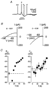
A, in order to determine the I-V relation of the glycine-induced current, voltage ramp protocols (Vh, −60 mV; range, −100 to 0 mV; duration, 100 ms) were applied before (con) and during the current response activated by 1 mm glycine (a-d). Note, current responses to voltage ramps are truncated due to the slow time resolution of the pen writer. B, I-V relations of the current responses corresponding to the voltage ramp responses a and c in A were created by subtracting the current response before glycine from the response obtained during the glycine action. The reversal potential during the outward current component (a - con) was −88 mV and during the inward current component (c - con) −52 mV, indicating that Egly was shifted about +36 mV during one minute. Traces are illustrated for the potential range between −100 and −40 mV. C, left panel, plot of Egly as a function of time during a glycine-evoked current response from a single neurone; □, cell membrane potential before glycine (−73 mV); ▪, Egly in the presence of glycine. Right panel, plot of mean Egly as a function of time obtained from 10 neurones. ^, mean membrane potential before glycine (−64 mV, s.d. 15 mV; n = 10); •, mean Egly in the presence of glycine; bars indicate s.d.; n = 2–10 for each data point; data were binned into classes of 6 s width. Dashed lines indicate the resting membrane potential. Note that Egly shifts on average by about 30 mV.
Figure 5
Figure 5. Glycine-induced tail currents in LSO neurones
In the presence of glycine (1 mm), tail currents were observed after the end of depolarizing rectangular command pulses (duration, 50 ms) when the voltage was stepped back to variable potentials (from −10 to −100 mV, as indicated above; Vh, −80 mV), and they lasted several seconds. The voltage protocols were applied in the absence and in the presence of glycine, giving rise to the tail currents illustrated in A and B, respectively. C, difference ‘B - A’ shows the net effect of glycine at different membrane potentials. Note that only the tail currents, but not the current responses to the preceding depolarizing rectangular command pulses are displayed in A-C. D, I-V relation. The peak of the tail current (indicated by an arrow in C) was plotted as a function of the voltage (reversal potential, −35 mV in this cell, but see text).
Figure 6
Figure 6. Effect of pulse duration (A) and pulse amplitude (B) on glycine-induced tail currents
A, five successive voltage steps (from Vh of −60 mV to +40 mV) with increasing duration (10, 50, 100, 150 and 200 ms) were applied. In the absence of glycine (a, control), the pulse duration had no effect on the tail current. With 1 mm glycine in the bath solution (b), a prolonged tail current component was present. The amplitude of the tail current increased with increasing pulse duration. Bottom traces show the subtracted current traces as indicated. B, three successive voltage steps (duration, 20 ms; Vh, −60 mV) to increasing levels (−20, +20 and +60 mV) were applied. In the absence of glycine (a, control), the pulse amplitude had no effect on the amplitude of the tail current. In the presence of 1 mm glycine (b), the amplitude of the tail current increased with increasing pulse amplitude. Bottom traces show the subtracted current traces. Pulse amplitudes are truncated and baselines were adjusted in A and B. Dotted line indicates zero current.
Figure 7
Figure 7. Coincident depolarizing activity shifts Egly in LSO neurones
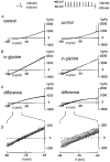
The coincident occurrence of spike activity and glycinergic input (left panel) was simulated by applying a train of 10 depolarizing pulses (from −80 mV to +40 mV; pulse duration 2 ms; pulse frequency 40 Hz; right panel) during the presence of currents activated by glycine (1 mm). Egly was determined using the voltage ramp method as described in the text. The time-dependent shift of Egly was determined by the successive application of 10 voltage ramps at 2 Hz. A, superposition of 10 current ramp responses in the absence of glycine (control). B, same as in A but in the presence of glycine. C, the current traces shown in A were subtracted from the corresponding traces displayed in B. The difference reflects the shift of Egly. D, part of C at higher magnification, illustrating the shift of the reversal potential.

Similar articles

Cited by

References

    1. Backus KH, Friauf E. Effects of synchronous depolarization on glycine-induced currents in developing rat auditory brainstem neurons. Society for Neuroscience Abstracts. 1996;22:647.
    1. Ballanyi K, Grafe P. An intracellular analysis of γ-aminobutyric-acid-associated ion movements in rat sympathetic ganglions. Journal of Physiology. 1985;365:41–58. - PMC - PubMed
    1. Barnes-Davis M, Forsythe ID. Pre- and postsynaptic glutamate receptors at a giant excitatory synapse in rat auditory brainstem slices. Journal of Physiology. 1995;488:387–406. - PMC - PubMed
    1. Ben-Ari Y, Cherubini E, Corradetti R, Gaiarsa JL. Giant synaptic potentials in immature rat CA3 hippocampal neurones. Journal of Physiology. 1989;416:303–325. - PMC - PubMed
    1. Bormann J, Hamill OP, Sakmann B. Mechanism of anion permeation through channels gated by glycine and gamma-aminobutyric acid in mouse cultured spinal neurones. Journal of Physiology. 1987;385:243–286. - PMC - PubMed

Publication types

MeSH terms

LinkOut - more resources