. 1998 Mar 15;507 ( Pt 3)(Pt 3):831-42.
doi: 10.1111/j.1469-7793.1998.831bs.x.

Interruption of central noradrenergic pathways and morphine withdrawal excitation of oxytocin neurones in the rat

Affiliations

Interruption of central noradrenergic pathways and morphine withdrawal excitation of oxytocin neurones in the rat

C H Brown et al. J Physiol. .

Abstract

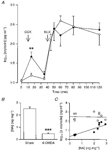
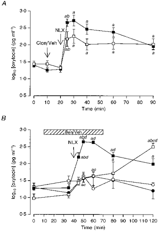
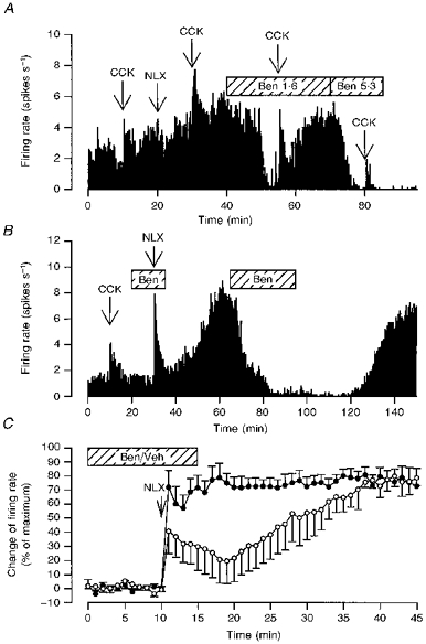
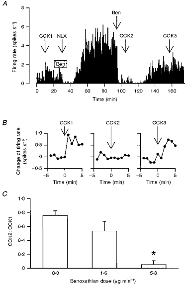
1. We have tested the hypothesis that morphine withdrawal excitation of oxytocin neurones that follows from administration of naloxone to morphine-dependent rats is a consequence of excitation of noradrenergic neurones. 2. Female rats were made morphine dependent by intracerebroventricular (i.c.v.) infusion of the opioid at increasing doses over 5 days. On the sixth day, the rats were anaesthetized with urethane or pentobarbitone and prepared for blood sampling to determine plasma oxytocin by radioimmunoassay or for in vivo extracellular recording of the firing rate of identified oxytocin neurones from the supraoptic nucleus. Morphine withdrawal was induced by intravenous (i.v.) injection of the opioid antagonist naloxone (5 mg kg-1). 3. In one group of rats the noradrenergic projections to the hypothalamus were lesioned by i.c.v. injection of 6-hydroxydopamine immediately prior to the induction of morphine dependence. In these rats the oxytocin secretion induced by i.v. cholecystokinin was reduced to 9 % of that seen in sham-lesioned rats but in contrast, no attenuation of morphine withdrawal-induced oxytocin secretion was observed. 4. i.c.v. infusion of the alpha1-adrenoreceptor antagonist benoxathian, at up to 5.3 microg min-1, dose- dependently inhibited the withdrawal excitation of oxytocin neurones in morphine-dependent rats under urethane anaesthesia, and benoxathian reduced withdrawal-induced oxytocin secretion to 37 % of that of vehicle-infused rats. i.c.v. benoxathian also inhibited the activity of oxytocin neurones in morphine-naïve rats. Similarly, microdialysis administration of 2 mM benoxathian directly onto the surface of the supraoptic nucleus reduced the activity of oxytocin neurones by 53 %. 5. Thus noradrenergic systems are not essential for the expression of morphine withdrawal excitation, since chronic neurotoxic destruction of the noradrenergic inputs to the hypothalamus did not affect the magnitude of withdrawal-induced oxytocin secretion. However, tonically active noradrenergic inputs influence the excitability of oxytocin neurones, and acute antagonism of this noradrenergic tone can powerfully impair the ability of oxytocin neurones to exhibit morphine withdrawal excitation.


Figures

Figure 1
Figure 1. Effects of chronic noradrenaline depletion on morphine withdrawal-induced hypersecretion of oxytocin
A, log10 plasma oxytocin concentrations (± s.e.m.) versus time in pentobarbitone-anaesthetized 6-OHDA- (○, n = 7) or sham-lesioned (•, n = 7) morphine-dependent rats administered CCK (20 μg kg−1, i.v.) at t = 10 min and naloxone (NLX; 5 mg kg−1, i.v.) at t = 40 min. Two-way repeated measures (RM) ANOVA showed that there was no effect of 6-OHDA lesion on the morphine-withdrawal hypersecretion of oxytocin. However, the change in oxytocin concentration following CCK was significantly reduced with respect to that in sham-lesioned rats (**P < 0.01, Mann-Whitney U test). B, hypothalamic noradrenaline concentrations (ng (mg wet weight)−1 ± s.e.m.) in sham- and 6-OHDA-lesioned rats at t = 120 min; ***P < 0.001, unpaired t test. C, scatter plot of the change (Δ) of (log10) plasma oxytocin concentration in individual sham- or 6-OHDA-lesioned, morphine-dependent rats following systemic CCK (20 μg kg−1, i.v.; •) or naloxone (5 mg kg−1, i.v.; ^) versus hypothalamic noradrenaline (NA) content (ng (mg wet weight)−1). There was a significant correlation of hypothalamic noradrenaline content with the change in oxytocin concentration after systemic CCK (r = 0.830; P = 0.002; n = 11), but not with that after naloxone (r = −0.053; P = 0.870; n = 12).
Figure 2
Figure 2. Effects of blockade of endogenous noradrenaline on morphine withdrawal-induced hypersecretion of oxytocin
A, plasma oxytocin concentrations (mean log10 plasma oxytocin concentration ±s.e.m.) measured in urethane-anaesthetized morphine-dependent rats. Morphine withdrawal was induced by injection of naloxone (NLX, 5 mg kg−1, i.v.). Rats were given an i.v. injection of either clonidine (Clon; 2.5 mg kg−1; □) or vehicle (Veh; 0.9% saline, ▪) 10 min before injection of naloxone. The increase in oxytocin concentration after naloxone was significantly less in the clonidine-treated rats (P < 0.05); two-way RM ANOVA revealed significant time (P < 0.0001) and interaction (P < 0.01) effects. a, P < 0.05 compared with basal concentration (t = 0 min); b, P < 0.05 compared with the preceding sample. B, plasma oxytocin concentrations (mean log10 plasma oxytocin concentration ±s.e.m.) measured in pentobarbitone-anaesthetized morphine-dependent rats (squares) and morphine-naïve rats (circles). Morphine withdrawal was elicited acutely by injection of naloxone (NLX, 5 mg kg−1, i.v.). Rats were given an i.c.v. infusion (hatched bar) of either benoxathian (5.3 μg min−1, i.c.v. at 0.53 μl min−1, open symbols) or vehicle (0.9% saline, closed symbols). Naloxone evoked an increased release of oxytocin in all groups (n = 6–8), but the magnitude of release was much greater in morphine-dependent rats. Benoxathian infusion attenuated and delayed the naloxone-induced secretion of oxytocin in dependent rats. Two-way ANOVA revealed a significant effect of morphine pretreatment (P < 0.0001) and a significant interaction between the effects of morphine pretreatment and benoxathian treatment (P < 0.05). Two-way RM ANOVA between the groups revealed significant group, time and interaction effects (all P < 0.0001). a, P < 0.05 compared with basal concentration (t = 0 min); b, P < 0.05 compared with the preceding sample; c, P < 0.05 compared with the appropriate time-matched, vehicle-treated, morphine-dependent controls; d, P < 0.05 compared with the similarly treated time-matched, morphine-naïve controls (Student-Newman-Keuls post hoc analyses).
Figure 3
Figure 3. Effects of acute α1-adrenoceptor antagonism on the activity of oxytocin neurones in morphine-naïve and morphine-dependent rats
A, the firing rate (averaged in consecutive 10 s intervals) of a single oxytocin cell recorded from a morphine-naïve rat administered CCK (20 μg kg−1, i.v.) at 10, 30, 55 and 80 min, naloxone (NLX, 5 mg kg−1, i.v.) at 20 min and infused i.c.v. with benoxathian at 1.6 μg min−1 (Ben 1.6) from 40 to 70 min and at 5.3 μg min−1 (Ben 5.3) from 70 to 85 min. Benoxathian inhibited spontaneous activity and attenuated the response to CCK. B, the firing rate (averaged in consecutive 10 s intervals) of a single oxytocin cell recorded from a morphine-dependent rat administered CCK (20 μg kg−1, i.v.) at 10 min, naloxone (NLX, 5 mg kg−1, i.v.) at 30 min and infused i.c.v. with benoxathian (Ben; 5.3 μg min−1) from 20 to 35 and 65 to 95 min. The first infusion of benoxathian delayed the peak sustained increase in firing rate after naloxone and a second infusion later reversibly inhibited the withdrawal excitation. C, effects of benoxathian on subsequent withdrawal excitation of oxytocin neurones. The composite change in firing rate of oxytocin neurones was induced by naloxone (NLX; 5 mg kg−1, i.v. at t = 10 min) in morphine-dependent rats during i.c.v. infusion of benoxathian (Ben; 5.3 μg min−1, n = 7, ^) or vehicle (Veh, n = 6, •). The change in firing rate was calculated for each cell by subtracting the initial mean basal firing rate (measured over 5 min in 1 min bins) from the firing rate measured in each minute, and was expressed as a percentage (mean ±s.e.m.) of the maximum change observed for each cell. The magnitude of withdrawal excitation was significantly lower in the first 5 min after naloxone in the benoxathian-infused group than in the vehicle-infused group (P < 0.05, two-way RM ANOVA followed by Student-Newman-Keuls analyses). In vehicle-infused rats, the naloxone-induced increase in firing rate was maximal within 5 min. In contrast, in benoxathian-infused rats the maximum firing rate was not attained until 30–35 min after naloxone, 25–30 min after the end of the benoxathian infusion (P < 0.05 compared with the 5 min immediately following naloxone administration to i.c.v. benoxathian-infused rats).
Figure 4
Figure 4. Effects of acute α1-adrenoceptor antagonism on CCK-induced excitation of oxytocin neurones
A, the firing rate of an oxytocin cell (averaged in consecutive 10 s intervals) in a morphine-dependent rat under urethane anaesthesia administered CCK (20 μg kg−1, i.v.), and infused i.c.v. with benoxathian (5.3 μg min−1, hatched box, Ben), naloxone (NLX; 5 mg kg−1, i.v.) and benoxathian (25 μg, i.c.v., Ben); the benoxathian infusion delayed the peak withdrawal response (see Fig. 3C) and the later injection of benoxathian reversibly inhibited the withdrawal excitation and the response to CCK. B, the change of firing rate of the oxytocin cell in A following each CCK injection (at t = 0 min) on an expanded time scale; the later injection of benoxathian blocked the response to i.v. CCK (CCK2). C, CCK2:CCK1 ratios from morphine-withdrawn and naïve rats treated with 0.3 (n = 3), 1.6 (n = 11) and 5.3 μg min−1 (n = 6) benoxathian i.c.v.; *P < 0.05 compared with the groups administered the lower two doses, one-way RM ANOVA followed by Student-Newman-Keuls analyses.

Similar articles

Cited by

References

    1. Aghajanian GK. Tolerance of locus coeruleus neurones to morphine and suppression of withdrawal response by clonidine. Nature. 1978;276:186–188. - PubMed
    1. Aghajanian GK, Kogan JH, Moghaddam B. Opiate withdrawal increases glutamate and aspartate efflux in the locus coeruleus: an in vivo microdialysis study. Brain Research. 1994;636:126–130. 10.1016/0006-8993(94)90186-4. - DOI - PubMed
    1. Akaoka H, Aston-Jones G. Opiate withdrawal-induced hyperactivity of locus coeruleus neurons is substantially mediated by augmented excitatory amino acid input. Journal of Neuroscience. 1991;11:3830–3839. - PMC - PubMed
    1. Bicknell RJ. Optimizing release from peptide hormone secretory nerve terminals. Journal of Experimental Biology. 1988;139:51–65. - PubMed
    1. Bicknell RJ, Leng G, Lincoln DW, Russell JA. Naloxone excites oxytocin neurones in the supraoptic nucleus of lactating rats after chronic morphine treatment. Journal of Physiology. 1988;396:297–317. - PMC - PubMed

Publication types

MeSH terms

LinkOut - more resources