. 1998 Mar;180(6):1586-91.
doi: 10.1128/JB.180.6.1586-1591.1998.

The folate branch of the methionine biosynthesis pathway in Streptomyces lividans: disruption of the 5,10-methylenetetrahydrofolate reductase gene leads to methionine auxotrophy

Affiliations

The folate branch of the methionine biosynthesis pathway in Streptomyces lividans: disruption of the 5,10-methylenetetrahydrofolate reductase gene leads to methionine auxotrophy

J Blanco et al. J Bacteriol. 1998 Mar.

Abstract

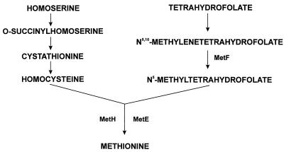
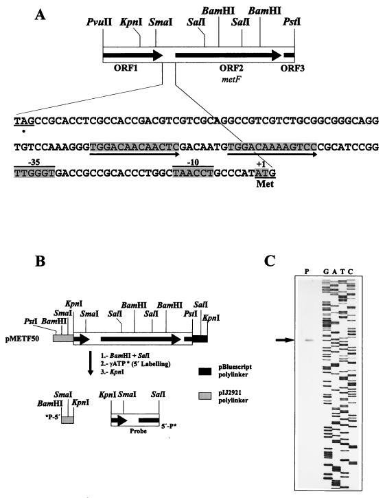
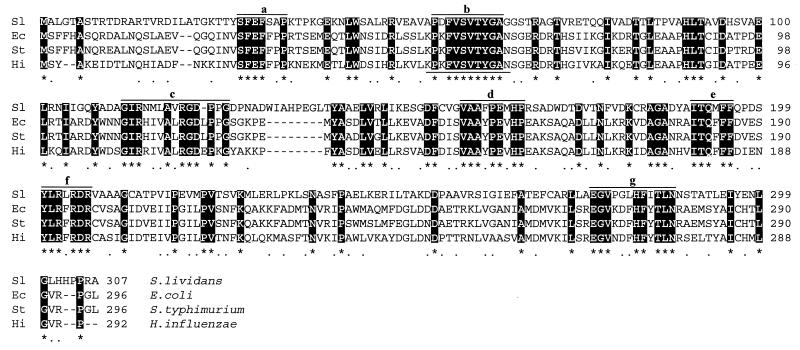
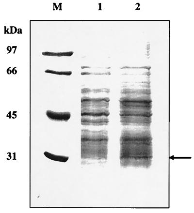
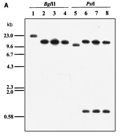
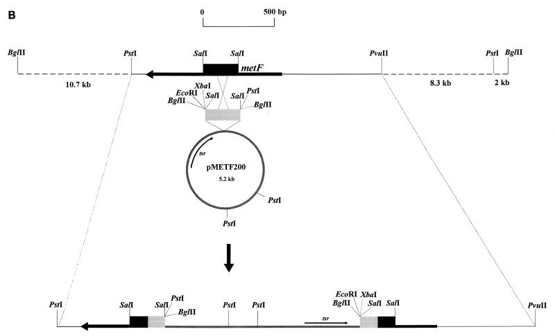
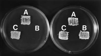
In enterobacteria, the methyl group of methionine is donated by 5-methyltetrahydrofolate that is synthesized from N5,10-methylenetetrahydrofolate by the 5,10-methylenetetrahydrofolate reductase. The Streptomyces lividans metF gene, which encodes 5,10-methylenetetrahydrofolate reductase, has been cloned. It encodes a protein of 307 amino acids with a deduced molecular mass of 33,271 Da. S1 exonuclease mapping of the transcription initiation site showed that the metF gene is expressed, forming a leaderless mRNA. A 13-bp tandem repeat located immediately upstream of the promoter region shows homology with the consensus MetR-binding sequence of Salmonella typhimurium. Expression of metF in multicopy plasmids in S. lividans resulted in accumulation of a 32-kDa protein, as shown by sodium dodecyl sulfate-polyacrylamide gel electrophoresis. Disruption of the metF gene led to methionine auxotrophy. Integration of the disrupting plasmid at the metF locus was confirmed by Southern hybridization in three randomly isolated transformants. The methionine auxotrophy was complemented by transformation of the auxotrophs with an undisrupted metF gene. These results indicate that the folate branch is essential for methionine biosynthesis in streptomycetes, as occurs in enterobacteria.


Figures

FIG. 1
FIG. 1
Biosynthetic pathway of methionine showing the formation of the homocysteine moiety from homoserine (left branch) and the origin of the methyl group from the folate branch (right branch). metF encodes the 5,10-methylenetetrahydrofolate reductase (MetF); MetE and MetH are alternative methyltransferases.
FIG. 2
FIG. 2
(A) Physical map of the 1.7-kb PvuII-PstI DNA region of S. lividans containing the metF gene (ORF2). The first in-frame ATG codon of ORF2 is underlined and labelled “Met.” The transcription start point is shaded and labelled +1, and the −10 and −35 boxes of the promoter region are indicated. The 13-bp direct repeat (putative MetR-binding site) is underlined with arrows. (B) Strategy for high-resolution S1 nuclease protection studies of the transcription start point. (C) Protected band (arrow) on S1 mapping experiments.
FIG. 3
FIG. 3
Alignment of the amino acid sequences of the methylenetetrahydrofolate reductases of S. lividans (AJ001630), E. coli (P00394), S. typhimurium (P11003), and H. influenzae (P45208) by using the CLUSTAL program. Conserved amino acids are in white-on-black type. Motifs a to g are sequences conserved in all tetrahydrofolate reductases. The amino acid sequence used for constructing the degenerate probe is underlined.
FIG. 4
FIG. 4
SDS-PAGE (12% polyacrylamide) of crude extracts of S. lividans(pIJ699) (lane 1) and S. lividans(pMETF150) (lane 2). Cultures were grown in NMMP medium (16) supplemented with thiostrepton (5 μg/ml). Lane M, SDS-PAGE molecular weight standards (low range; Bio-Rad). Sizes are indicated on the left. The overexpressed protein is indicated with an arrow.
FIG. 5
FIG. 5
(A) Disruption of the S. lividans metF gene, as shown by hybridization with a 255-bp SalI probe internal to metF. Lanes: 1 and 5, undisrupted S. lividans 1326 (control); 2 and 6, S. lividans MD1 (met); 3 and 7, S. lividans MD2 (met); 4 and 8, S. lividans MD3 (met). Total DNA of each strain was digested with BglII (lanes 1 to 4) or PstI (lanes 5 to 8). (B) Disruption of metF by integration of pMETF200; tsr, thiostrepton resistance gene (used as a marker).
FIG. 5
FIG. 5
(A) Disruption of the S. lividans metF gene, as shown by hybridization with a 255-bp SalI probe internal to metF. Lanes: 1 and 5, undisrupted S. lividans 1326 (control); 2 and 6, S. lividans MD1 (met); 3 and 7, S. lividans MD2 (met); 4 and 8, S. lividans MD3 (met). Total DNA of each strain was digested with BglII (lanes 1 to 4) or PstI (lanes 5 to 8). (B) Disruption of metF by integration of pMETF200; tsr, thiostrepton resistance gene (used as a marker).
FIG. 6
FIG. 6
Growth in Streptomyces minimal medium of the parental strain S. lividans 1326 (A), the disrupted S. lividans MD1 (B), and a transformant of S. lividans MD1 with plasmid pVKK-metF (C). The righthand plate was supplemented with l-methionine (50 μg/ml).

References

    1. Anné J, Van Mellaert L. Streptomyces lividans as host for heterologous protein production. FEMS Microbiol Lett. 1993;114:121–128. - PubMed
    1. Belfaiza J, Parsot C, Martel A, Buothier de la Tour C, Margarita D, Cohen G N, Saint-Girons I. Evolution in the biosynthesis pathways: two enzymes catalyzing consecutive steps in methionine biosynthesis originate from a common ancestor and possess a similar regulatory region. Proc Natl Acad Sci USA. 1986;83:867–871. - PMC - PubMed
    1. Bibb M J, Cohen S N. Gene expression in Streptomyces: construction and application of promoter-probe plasmid vectors in Streptomyces lividans. Mol Gen Genet. 1982;187:265–277. - PubMed
    1. Chary, V. K., and J. F. Martín. Unpublished data.
    1. Coque J J R, Enguita F J, Martín J F, Liras P. A two-protein component 7α-cephem-methoxylase encoded by two genes of the cephamycin C cluster converts cephalosporin C to 7-methoxycephalosporin C. J Bacteriol. 1995;177:2230–2235. - PMC - PubMed

Publication types

MeSH terms

Associated data

LinkOut - more resources