

Abnormal mucus in cap polyposis
- PMID: 9518233
- PMCID: PMC1726938
- DOI: 10.1136/gut.42.1.135
Abnormal mucus in cap polyposis
Abstract
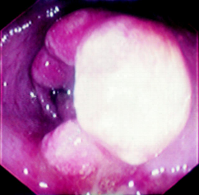
Background: Cap polyposis is a rare disease characterised by mucoid and bloody diarrhoea, with polyps covered by a cap of mucoid and fibrinopurulent exudate. The pathogenesis is not known.
Aims: To pour some light on cap polyposis pathogenesis, by examining the mucus of patients and analysing the expression of five mucin genes, MUC2, MUC3, MUC4, MUC5AC, and MUC5B.
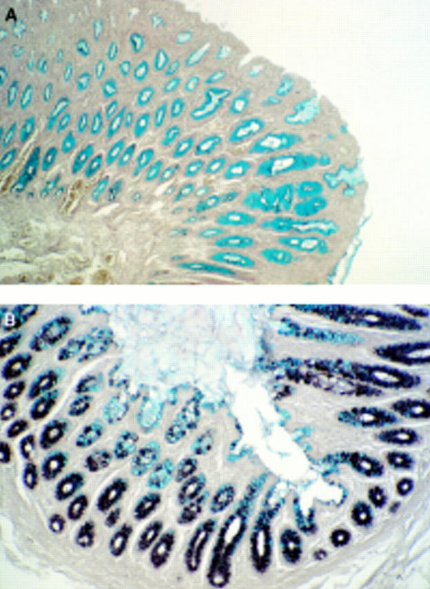
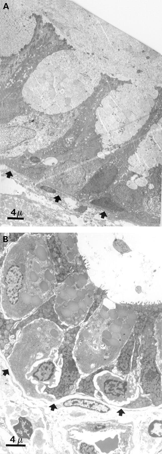
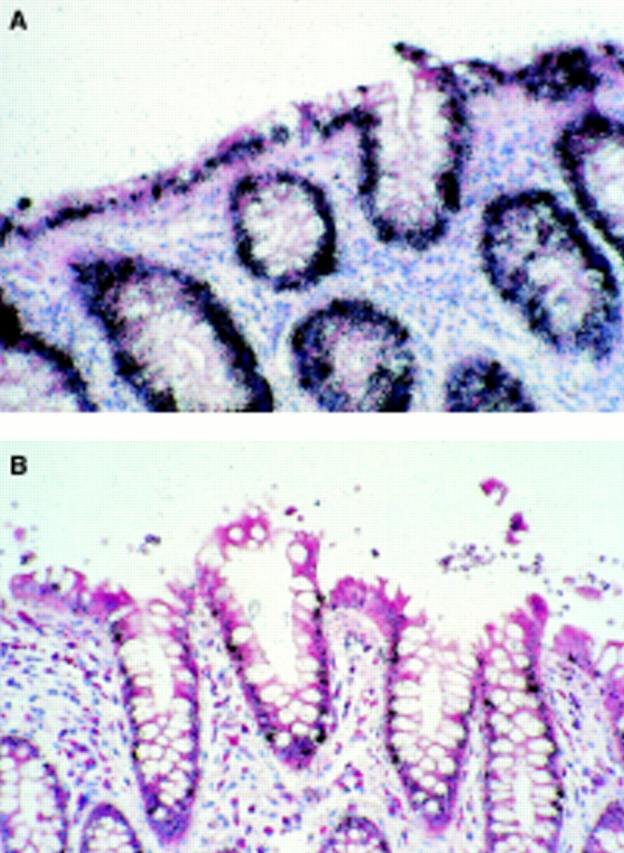
Patient and methods: The study was performed on biopsy specimens taken from a patient with recurrent cap polyposis. Histochemical examination, electron microscopy, and mRNA in situ hybridisation were used.
Results: The mucus of cap polyposis differed in three respects from that of normal adult colon: abnormal ultrastructure of the mucus in the goblet cells, predominance of non-sulphated mucins, abnormal expression of the MUC4, MUC3, and MUC5AC genes.
Conclusions: Most of these abnormalities have been reported for other pathological situations, suggesting that the abnormalities observed in the mucus of this patient with cap polyposis are probably secondary phenomena rather than primary. However, the mucin abnormalities detected, which reflect deregulation of the expression of three apomucin genes, abnormal glycosylation, and abnormalities of the secretion process, are also probably involved in the clinical manifestations of cap polyposis.
Figures
Publication types
MeSH terms
Substances
LinkOut - more resources
Full Text Sources
Medical
Research Materials
Miscellaneous