Review
. 1998 May 1;508 ( Pt 3)(Pt 3):647-57.
doi: 10.1111/j.1469-7793.1998.647bp.x.

Structure and function of voltage-gated sodium channels

Affiliations
Review

Structure and function of voltage-gated sodium channels

E Marban et al. J Physiol. .

Abstract

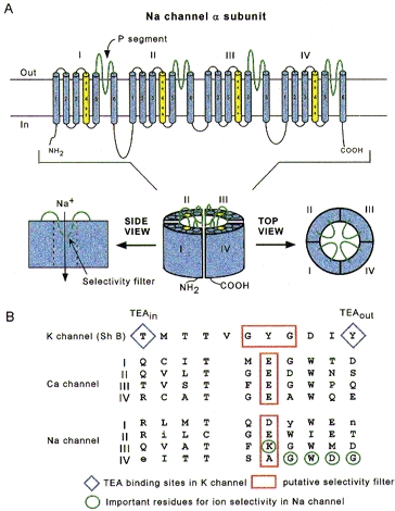
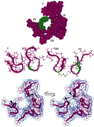
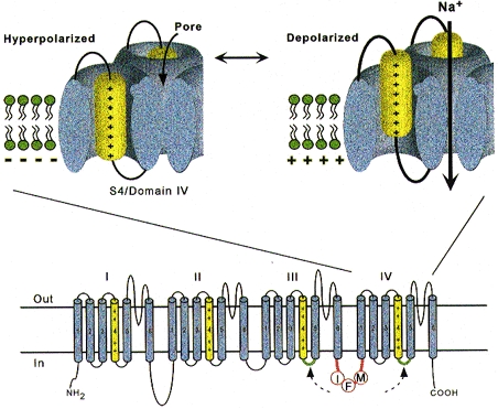
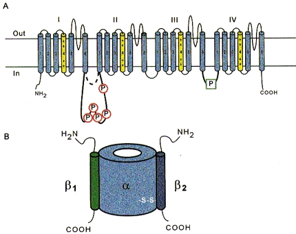
1. Sodium channels mediate fast depolarization and conduct electrical impulses throughout nerve, muscle and heart. This paper reviews the links between sodium channel structure and function. 2. Sodium channels have a modular architecture, with distinct regions for the pore and the gates. The separation is far from absolute, however, with extensive interaction among the various parts of the channel. 3. At a molecular level, sodium channels are not static: they move extensively in the course of gating and ion translocation. 4. Sodium channels bind local anaesthetics and various toxins. In some cases, the relevant sites have been partially identified. 5. Sodium channels are subject to regulation at the levels of transcription, subunit interaction and post-translational modification (notably glycosylation and phosphorylation).


Figures

Figure 1
Figure 1. Schematic depictions of the Na+ channel α subunit
A, putative transmembrane folding. The charged S4 segments are shown in yellow, and the pore-lining P segments in green. B, aligned primary amino acid sequences in single-letter code of the P segments in a K+ channel (Shaker B), the four domains of the cardiac L-type Ca2+ channel, and the four domains of the Na+ channel. Residues shown in upper case are highly conserved among voltage-dependent Na+ channels. The diamonds indicate the external and internal binding sites for tetraethylammonium (TEA) ion in the K+ channel and the red boxes outline the putative selectivity filters, although, in the case of the Na+ channel, the residues which are most important for selectivity (circled in green) are mostly outside the box.
Figure 2
Figure 2. Structure for the Na+ channel pore proposed by Bénitah et al. (1997)
Residues which play a particularly prominent role in determining Na+ selectivity are shown in green. Top: space-filling model of the Na+ channel pore consistent with the available mutagenesis data. Centre: side views of the P segments as they might appear to permeant ions, in ribbon format. Bottom: Na+ channel pore motions illustrated by the pairing of residues D400 and G1530 (arrowheads), which can form an internal disulphide in the pore when substituted with cysteines (Bénitah et al. 1997; Tsushima et al. 1997).
Figure 3
Figure 3. Schematic depictions of the Na+ channel α subunit illustrating the S4 activation sensors (top) and the III-IV linker which contributes to fast inactivation (bottom)
The putative receptors of the III-IV linker are shown in green. Modified from Yang et al. (1996).
Figure 4
Figure 4. Na+ channel phosphorylation sites and subunits
A, schematic diagram highlighting the prominent phosphorylation sites of the α subunit. These are in the III-IV linker, and in the long variant of the I-II linkers; the length of this linker is isoform specific. B, maximal subunit composition of the Na+ channel. Note that not all Na+ channels include either or both β subunits, but the α subunit is obligatory for function.

References

    1. Adelman WJ, Palti Y. The effects of external potassium and long duration voltage conditioning on the amplitude of sodium currents in the giant axon of the squid, Loligo pealei. Journal of General Physiology. 1969;54:589–606. - PMC - PubMed
    1. Aldrich RW, Corey DP, Stevens CF. A reinterpretation of mammalian sodium channel gating based on single channel recording. Nature. 1983;306:436–441. - PubMed
    1. Backx P, Yue D, Lawrence J, Marban E, Tomaselli G. Molecular localization of an ion-binding site within the pore of mammalian sodium channels. Science. 1992;257:248–251. - PubMed
    1. Balser J, Nuss H, Chiamvimonvat N, Pérez-García M, Marban E, Tomaselli G. External pore residue mediates slow inactivation in μ1 rat skeletal muscle sodium channels. The Journal of Physiology. 1996a;494:431–442. - PMC - PubMed
    1. Balser J, Nuss H, Orias D, Johns D, Marban E, Tomaselli G, Lawrence J. Local anesthetics as effectors of allosteric gating. Lidocaine effects on inactivation-deficient rat skeletal muscle Na+ channels. Journal of Clinical Investigation. 1996b;98:2874–2886. - PMC - PubMed

Publication types

Substances

LinkOut - more resources