. 1998 Feb 15;6(2):135-45.
doi: 10.1016/s0969-2126(98)00016-1.

Structural analysis of the Spiroplasma virus, SpV4: implications for evolutionary variation to obtain host diversity among the Microviridae

Affiliations

Structural analysis of the Spiroplasma virus, SpV4: implications for evolutionary variation to obtain host diversity among the Microviridae

P R Chipman et al. Structure. .

Abstract

Background: Spiroplasma virus, SpV4, is a small, non-enveloped virus that infects the helical mollicute Spiroplasma melliferum. SpV4 exhibits several similarities to the Chlamydia phage, Chp1, and the Coliphages alpha 3, phi K, G4 and phi X174. All of these viruses are members of the Microviridae. These viruses have isometric capsids with T = 1 icosahedral symmetry, cause lytic infections and are the only icosahedral phages that contain single-stranded circular DNA genomes. The aim of this comparative study on these phages was to understand the role of their capsid proteins during host receptor recognition.

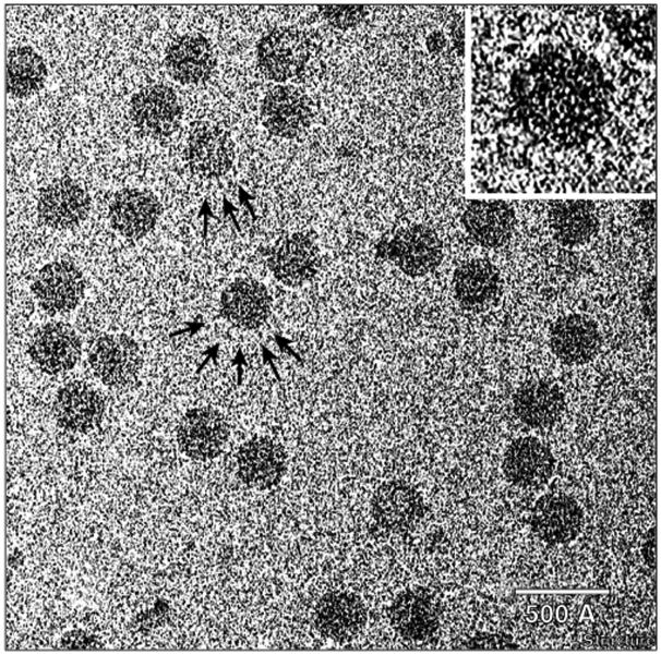
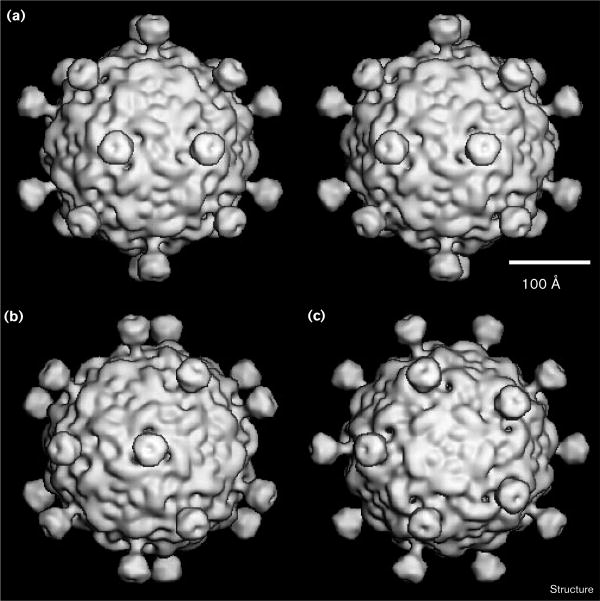
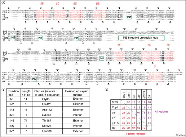
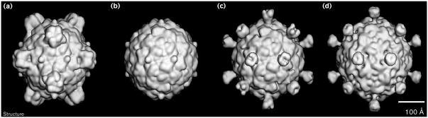
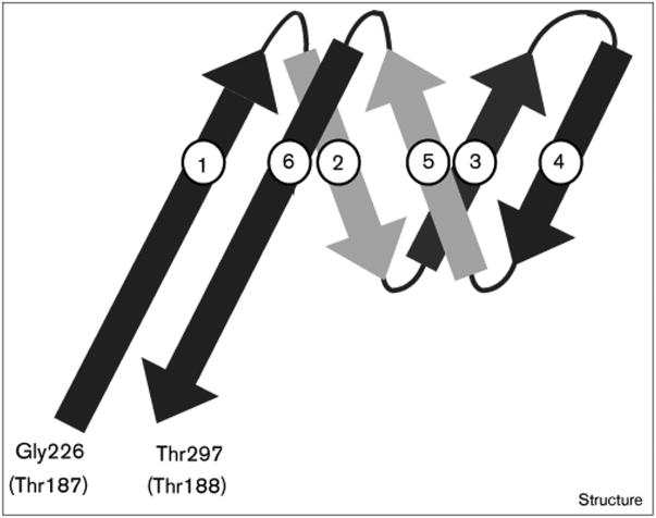
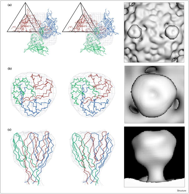
Results: The three-dimensional structure of SpV4 was determined to 27 A resolution from images of frozen-hydrated particles. Cryo-electron microscopy (cryo-EM) revealed 20, approximately 54 A long, 'mushroom-like' protrusions on the surface of the capsid. Each protrusion comprises a trimeric structure that extends radially along the threefold icosahedral axes of the capsid. A 71 amino acid portion of VP1 (the SpV4 capsid protein) was shown, by structural alignment with the atomic structure of the F capsid protein of phi X174, to represent an insertion sequence between the E and F strands of the eight-stranded antiparallel beta-barrel. Secondary structure prediction of this insertion sequence provided the basis for a probable structural motif, consisting of a six-stranded antiparallel beta sheet connected by small turns. Three such motifs form the rigid stable trimeric structures (mushroom-like protrusions) at the threefold axes, with hydrophobic depressions at their distal surface.

Conclusions: Sequence alignment and structural analysis indicate that distinct genera of the Microviridae might have evolved from a common primordial ancestor, with capsid surface variations, such as the SpV4 protrusions, resulting from gene fusion events that have enabled diverse host ranges. The hydrophobic nature of the cavity at the distal surface of the SpV4 protrusions suggests that this region may function as the receptor-recognition site during host infection.


Figures

Figure 1
Figure 1
SpV4 particles negatively stained with 1% uranyl acetate on a carbon support film. Arrows highlight protrusions on three particles. The inset, a close-up view of one particle, shows several, stain-excluding (white) ‘dots’ inside the particle. The dots are likely to be end-on views of the protrusions. The scale bar = 500 Å.
Figure 2
Figure 2
Ribbon drawing of the atomic structure of the φX174 capsid protein F. The β-barrel motif is coloured red with the β strands (B, I, D, G, and C, H, E, F) labeled according to standard convention. Arrows highlight the equivalent positions of the capsid protein VP1 of SpV4, where insertion loops (identified as IN1–7) occur relative to the capsid protein F of φX174 (refer to Figure 6). Residue numbering is given for the F protein of φX174. The orientation of the F protein is shown viewed towards the interior of the virus down a twofold axis. An icosahedral asymmetric unit (large open triangle) includes the region bounded by a fivefold axis (filled pentagon) and two adjacent threefold axes (filled triangles). (Figure produced with MOLSCRIPT [53].)
Figure 3
Figure 3
Unstained SpV4 particles suspended in a layer of vitreous ice over holes in a carbon support film. Arrows highlight several protrusions on two particles. The inset shows an enlarged view of one of the highlighted particles. The scale bar = 500 Å.
Figure 4
Figure 4
Shaded surface representations of the SpV4 three-dimensional reconstruction. The representations are viewed down the (a) twofold (in stereo), (b) threefold and (c) fivefold axes. The reconstruction was computed to 27 Å resolution from 22 different SpV4 particle images. The scale bar = 100 Å.
Figure 5
Figure 5
Close-up views of one SpV4 trimeric protrusion. (a) Shaded surface view from above the top of the protrusion. (b) Side view, as from the bottom of (a), with the front of the map (as indicated by the dotted line towards the bottom of (a)) removed to reveal a channel near the base of the stalk. (c) The same view as (b) but with more density removed, to dashed line in center of (a), to reveal a cross-section through the center of the protrusion. (d) Density distribution in the center of the protrusion at the same plane where the map was cut in (c). Highest density features (protein) appear black, whereas lowest densities (solvent) appear white or gray. This representation gives a more realistic rendering of the density fluctuations inside the protrusion and, for example, shows a low density along the vertical axis at the top of the protrusion. (e) The view is the same as in (c) but with four density contour levels shown to illustrate the varying density in the cross-section.
Figure 6
Figure 6
Comparison of the amino acid sequences of capsid proteins of members of the Microviridae family. (a) Sequence alignment of VP1 of SpV4 and Chp1 and protein F for the Coliphages α3, φK, φX174 and G4. Bold letters indicate residues that are completely conserved in all the phages compared; residues that form part of the β-barrel structure motif elements are colored red and labeled (as shown in Figure 2). The sequence numbers are given for SpV4 (top) and φX174 (bottom). Insertions are labeled and marked by green boxes. (b) Assignment and location of the seven insertion loops (assigned as IN1–7) found in the SpV4 and Chp1 sequences in relation to the Coliphages. (c) The percentage of identical amino acids between the aligned capsid proteins, for all residues (shown in purple) and those involved in the β-barrel motif (shown in red).
Figure 7
Figure 7
Shaded surface representation comparisons of φX174 and SpV4 structures. (a) The atomic φX174 structure [14]. (b) The φX174 structure, as in (a), but with the prominent spikes (G protein pentamers) removed to reveal just the F capsid. (c) An SpV4–φX174 hybrid model, formed by combining the φX174 F capsid model (b) with the pseudo-atomic model of the SpV4 protrusion. (d) The cryo-EM reconstruction of SpV4. All structures are shown at ∼27 Å resolution and are viewed along a twofold axis of symmetry. The scale bar = 100 Å.
Figure 8
Figure 8
Phylogenetic tree for six Microviridae capsid proteins.
Figure 9
Figure 9
Proposed structural motif model for residues Gly226–Thr297 of the capsid protein of SpV4. These residues comprise one third of the protrusion at each threefold axis. The residues in the F protein of φX174 at the exit and entry points for the SpV4 loop are indicated in parentheses; β strands are shown as arrows.
Figure 10
Figure 10
Stereo views of the atomic model of the SpV4 protrusion. (a) The fit of a pseudo-atomic model of an SpV4 VP1 trimer (separate monomers colored blue, green and red) into the cryo-EM reconstruction (gray isodensity contour). Shown also is the viral asymmetric unit, depicted as an open triangle. The view is along a twofold axis. The fit of three symmetry-related SpV4 loop structures (Gly226–Thr297) into the cryo-EM density map, as viewed from (b) the top, along a threefold axis or (c) the side, perpendicular to a threefold axis. Corresponding shaded surface representations are shown to the right of each stereo view. (The stereo views were produced with MacInPlot [54].)

References

    1. Ricard B, Garnier M, Bove JM. Characterization of SPV3 from spiroplasmas and discovery of a new spiroplasma virus (SPV4) Rev Infect Dis. 1982;4:S275.
    1. Renaudin J, Pascarel MC, Garnier M, Carle P, Bove JM. Characterization of spiroplasma virus group 4 (SPV4) Isr J Med Sci. 1984;20:797–799. - PubMed
    1. Renaudin J, Pascarel MC, Garnier M, Junca-Carle P, Bove JM. SpV4, a new spiroplasma virus with circular, single stranded DNA. Ann Virol. 1984;135E:343–361.
    1. Woese CR. Bacterial evolution. Microbiol Rev. 1987;51:221–271. - PMC - PubMed
    1. Weisburg WG, et al. Woese CR. A phylogenetic analysis of the mycoplasmas: basis for their classification. J Bacteriol. 1989;171:6455–6467. - PMC - PubMed

Publication types

Associated data

LinkOut - more resources