

Expression of the somatostatin subtype 2A receptor in the rabbit retina
- PMID: 9520104
- PMCID: PMC3696039
- DOI: 10.1002/(sici)1096-9861(19980330)393:1<93::aid-cne9>3.0.co;2-l
Expression of the somatostatin subtype 2A receptor in the rabbit retina
Abstract
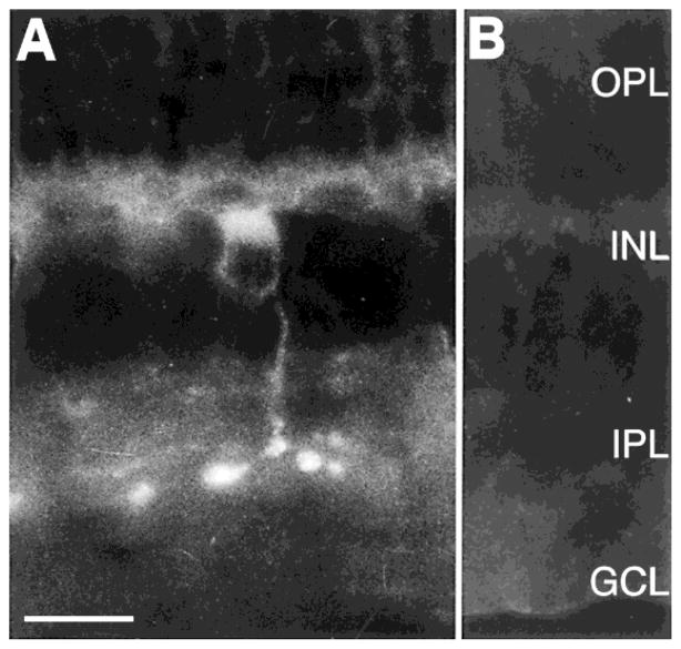
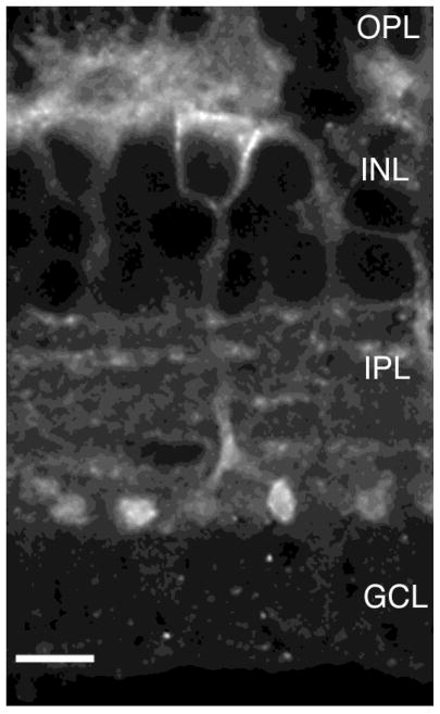
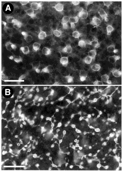
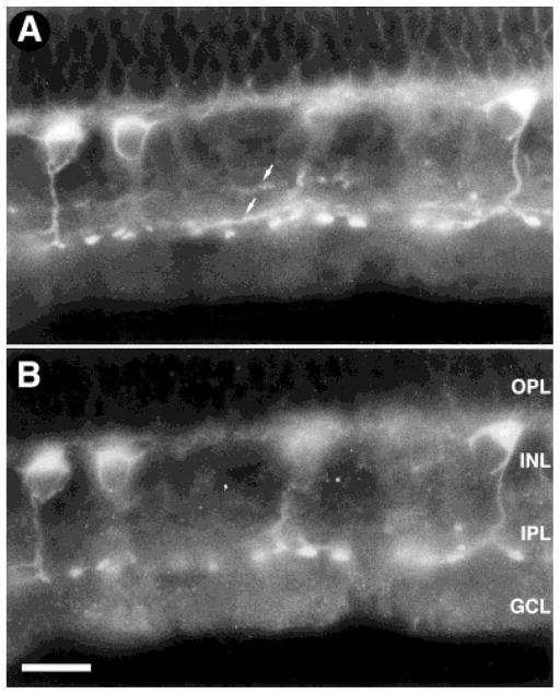
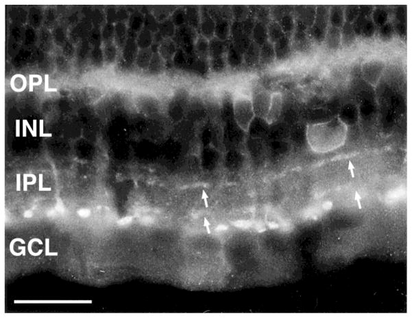
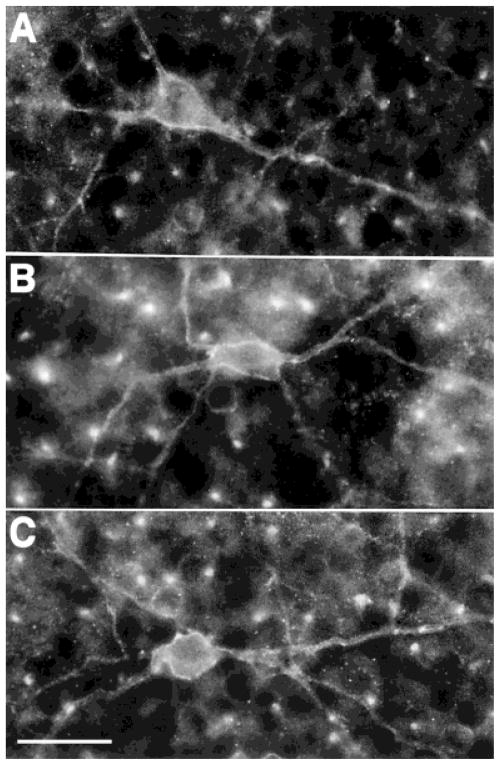
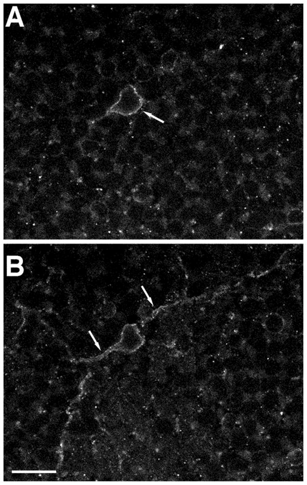
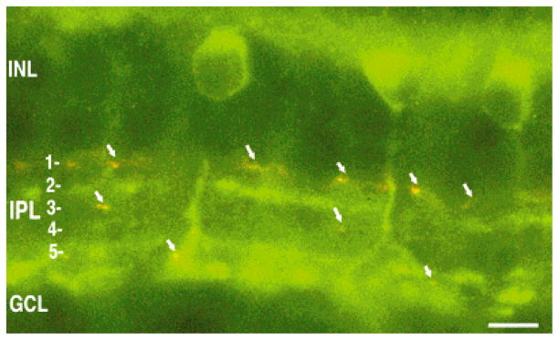
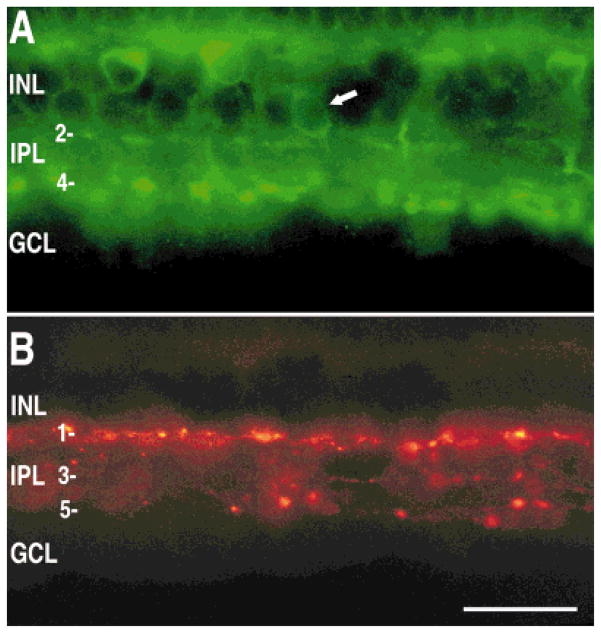
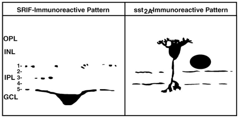
In the retina, somatostatin influences neuronal activity likely by acting at one or more somatostatin subtype (sst) receptors. Somatostatin and somatostatin-binding sites are distributed predominantly to the inner retina. The present study has investigated the cellular expression of one of the sst receptors, the sst2A receptor isoform, in the rabbit retina. These studies have used a new polyclonal antibody directed to the predicted C-terminus of mouse sst2A(361-369) receptor. Antibody specificity was tested by preadsorption of the primary antibody with a peptide corresponding to sst2A(361-369). sst2A Receptor immunoreactivity was localized mainly to the plasma membrane of rod bipolar cells and to sparsely occurring, wide-field amacrine cells. Immunostaining in rod bipolar cells was strongest in the axon and axon terminals in lamina 5 of the inner plexiform layer (IPL) and was weakest in the cell body and dendrites. Double-labeling experiments using a monoclonal antibody against protein kinase C (PKC; alpha and beta), a rod bipolar cell-selective marker, showed complete colocalization. In horizontal sections of retina, immunostained bipolar cell bodies had a dense distribution, which is in agreement with the reported distribution of rod bipolar cell bodies. Immunoreactive amacrine cell bodies were located at the border of the inner nuclear layer and the IPL, and thin varicose processes ramified mainly in laminae 2 and 4 of the IPL. These observations indicate that somatostatin influences visual information processing in the retina 1) by acting presynaptically on rod bipolar cell axon terminals and b) by influencing the activity of sparsely occurring amacrine cells.
Figures
Similar articles
-
Somatostatin receptor subtype 2A expression in the rat retina.Neuroscience. 1999;94(3):675-83. doi: 10.1016/s0306-4522(99)00170-0. Neuroscience. 1999. PMID: 10579559
-
Somatostatin and somatostatin subtype 2A expression in the mammalian retina.Microsc Res Tech. 2000 Jul 15;50(2):103-11. doi: 10.1002/1097-0029(20000715)50:2<103::AID-JEMT2>3.0.CO;2-X. Microsc Res Tech. 2000. PMID: 10891874 Review.
-
Postnatal development of somatostatin 2A (sst2A) receptors expression in the rabbit retina.Brain Res Dev Brain Res. 2000 Sep 30;123(1):67-80. doi: 10.1016/s0165-3806(00)00073-0. Brain Res Dev Brain Res. 2000. PMID: 11020551
-
Somatostatin (SRIF) and SRIF receptors in the mouse retina.Brain Res. 2002 May 17;936(1-2):1-14. doi: 10.1016/s0006-8993(02)02450-2. Brain Res. 2002. PMID: 11988224
-
Localization of five somatostatin receptors in the rat central nervous system using subtype-specific antibodies.J Physiol Paris. 2000 May-Aug;94(3-4):259-64. doi: 10.1016/s0928-4257(00)00212-6. J Physiol Paris. 2000. PMID: 11088003 Review.
Cited by
-
Synaptic transmission at retinal ribbon synapses.Prog Retin Eye Res. 2005 Nov;24(6):682-720. doi: 10.1016/j.preteyeres.2005.04.002. Prog Retin Eye Res. 2005. PMID: 16027025 Free PMC article. Review.
-
Molecular and Cellular Mechanisms Underlying Somatostatin-Based Signaling in Two Model Neural Networks, the Retina and the Hippocampus.Int J Mol Sci. 2019 May 21;20(10):2506. doi: 10.3390/ijms20102506. Int J Mol Sci. 2019. PMID: 31117258 Free PMC article. Review.
-
Somatostatin modulates voltage-gated K(+) and Ca(2+) currents in rod and cone photoreceptors of the salamander retina.J Neurosci. 2000 Feb 1;20(3):929-36. doi: 10.1523/JNEUROSCI.20-03-00929.2000. J Neurosci. 2000. PMID: 10648697 Free PMC article.
-
Localization of neuropeptide Y1 receptor immunoreactivity in the rat retina and the synaptic connectivity of Y1 immunoreactive cells.J Comp Neurol. 2002 Dec 23;454(4):373-82. doi: 10.1002/cne.10423. J Comp Neurol. 2002. PMID: 12455004 Free PMC article.
-
Effect of somatostatin analogues on chemically induced ischaemia in the rat retina.Naunyn Schmiedebergs Arch Pharmacol. 2005 Jan;371(1):44-53. doi: 10.1007/s00210-004-1011-9. Epub 2005 Jan 12. Naunyn Schmiedebergs Arch Pharmacol. 2005. PMID: 15645293
References
-
- Ayoub GS, Matthews G. Substance P modulates calcium current in retinal bipolar neurons. Vis Neurosci. 1992;8:539–544. - PubMed
-
- Brazeau P, Vale W, Burgus R, Ling N, Butcher M, Rivier J, Guillemin R. Hypothalamic polypeptide that inhibits the secretion of immunoreactive pituitary growth hormone. Science. 1973;179:77–79. - PubMed
-
- Brecha NC, Casini G, Rickman D. Organization and development of sparsely distributed wide-field amacrine cells in the rabbit retina. In: Bagnoli P, Hodges W, editors. The Changing Visual System: Maturation and Aging in the Central Nervous System. London: Plenum Press; 1991. pp. 95–117.
Publication types
MeSH terms
Substances
Grants and funding
LinkOut - more resources
Full Text Sources
