. 1998 Mar 31;95(7):3425-30.
doi: 10.1073/pnas.95.7.3425.

The transmembrane domain in viral fusion: essential role for a conserved glycine residue in vesicular stomatitis virus G protein

Affiliations

The transmembrane domain in viral fusion: essential role for a conserved glycine residue in vesicular stomatitis virus G protein

D Z Cleverley et al. Proc Natl Acad Sci U S A. .

Abstract

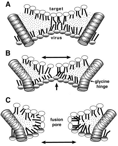
The transmembrane (TM) domains of viral fusion proteins are required for fusion, but their precise role is unknown. G protein, the fusion protein of vesicular stomatitis virus, was previously shown to lose syncytia-forming ability if six residues (GLIIGL) were deleted from its TM domain. The 20-residue TM domain of wild-type (TM20) G protein was thus changed into a TM domain of 14 residues (TM14). To assess possible sequence specificity for this loss of function, the two Gly residues in TM20 were replaced with either Ala or Leu. Both mutations resulted in complete loss of fusion activity, as measured by fusion-dependent reporter gene transfer. Single substitutions decreased activity by about half. TM14 was weakly active (15%) but reintroduction of a Gly residue into TM14 by a single Ile --> Gly substitution increased activity to 80%. All mutants retained normal hemifusion activity, i.e., lipid mixing between the outer leaflets of the reacting membranes. Thus, at least one TM Gly residue is required for a late step in fusion mediated by G protein. Gly residues were significantly (2.6-fold; P = 0.004) more abundant in the TM domains of viral fusion proteins than in those of nonfusion proteins and were distributed differently within the TM domain. Thus, Gly residues in the TM domain of other viral fusion proteins may also prove to be important for fusion activity.


Figures

Figure 1
Figure 1
Amino acid sequences of the TM domains of VSV G protein (Indiana serotype) and the mutants used in this study. The wild-type (TM20) sequence is shown, numbered from the cytoplasmic end (cyto), with the residues that were deleted to form TM14 (12) boxed.
Figure 2
Figure 2
Fusion activity of VSV G protein and of the mutants shown in Fig. 1, as determined by the reporter gene activation assay. Results are the mean and SD (error bars) of at least three experiments, each normalized to results with TM20, because absolute numbers varied. In each experiment the number of foci in at least two plates transfected with each construct were counted. Transfection efficiencies were uniformly 45–55%, in all experiments and for each construct. TM20 plates generally had 100–200 foci per plate. Background foci produced by pGEM-transfected cells lacking any G insert (usually 10–20% of TM20 levels) were subtracted from all values.
Figure 3
Figure 3
R18 dye transfer induced by low pH from dye-loaded High Five cells into HeLa cells expressing the following constructs: (A) pGEM alone. (B) TM20. (C) TM14. (D) A6,10. (E) L6,10. R18 transfer, indicative of lipid mixing, has occurred in all cells except those in A. (×200.)
Figure 4
Figure 4
Lucifer yellow dye transfer induced by low pH from dye-loaded High Five cells into HeLa cells expressing the following constructs: (A) pGEM alone. (B) TM20. (C) TM14. (D) A6,10. (E) L6,10. Lucifer yellow transfer, indicative of aqueous compartment mixing, has occurred only in cells in B. (×400.)
Figure 5
Figure 5
Frequency of occurrence of Gly residues in specific locations within the TM domains of fusion and nonfusion proteins listed in Tables 1 and 2. Location intervals are numbered from the cytoplasmic end of the TM domain as shown in Fig. 1.
Figure 6
Figure 6
Model for the participation of a TM Gly residue in fusion. (A) Rigid TM domains are embedded in the viral (lower) membrane and positioned around a hemifusion diaphragm, composed of the inner leaflets of the two reacting membranes. Because hemifusion has already occurred, a single outer leaflet (not shown) surrounds the structure shown in three dimensions. (B) Same as A, but with TM domain containing a bend around a Gly “hinge.” This could result in compression (increased negative curvature) of the viral leaflet and expansion of the target (upper) leaflet of the hemifusion diaphragm. (C) The perturbed lipids rearrange to form a more stable fusion pore. Expansion occurs in the direction indicated by the arrows.

References

    1. Kemble G W, Danieli T, White J M. Cell. 1994;76:383–391. - PubMed
    1. Melikyan G B, White J M, Cohen F S. J Cell Biol. 1995;131:679–691. - PMC - PubMed
    1. Melikyan G B, Brener S A, Ok D C, Cohen F S. J Cell Biol. 1997;136:995–1005. - PMC - PubMed
    1. Ragheb J A, Anderson F. J Virol. 1994;68:3207–3219. - PMC - PubMed
    1. Salzwedel K, Johnston P B, Roberts S J, Dubay J W, Hunter E. J Virol. 1993;67:3264–3273. - PMC - PubMed

LinkOut - more resources