. 1998 Mar 31;95(7):3615-20.
doi: 10.1073/pnas.95.7.3615.

Identification of cytokeratin 1 as a binding protein and presentation receptor for kininogens on endothelial cells

Affiliations

Identification of cytokeratin 1 as a binding protein and presentation receptor for kininogens on endothelial cells

A A Hasan et al. Proc Natl Acad Sci U S A. .

Abstract

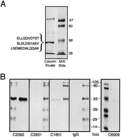
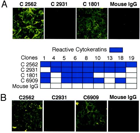
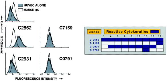
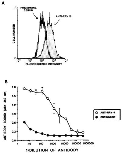
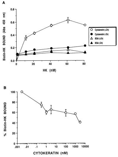
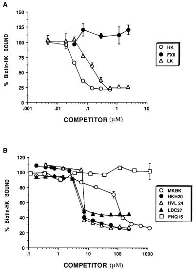
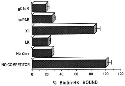
A kininogen binding protein(s), a putative receptor, was identified on endothelial cells. A 54-kDa protein was isolated by a biotin-high molecular mass kininogen (HK) affinity column that, on aminoterminal sequencing of tryptic digests, was identified as cytokeratin 1. Multiple antibodies directed to cytokeratin 1 reacted with a 54-kDa band on immunoblot of lysates of endothelial cells. On laser scanning confocal microscopy, cytokeratin 1 antigen was found on the surface of endothelial cells. Cytokeratin 1 antigen also was detected on endothelial cell membranes by flow cytometry. Moreover, an antipeptide antibody to a sequence unique to cytokeratin 1 also specifically bound to nonpermeabilized endothelial cells. Biotin-HK specifically bound to cytokeratin only in the presence of Zn2+, and cytokeratin blocked biotin-HK binding to endothelial cells. Further, HK and low molecular mass kininogen, but not factor XII, blocked biotin-HK binding to cytokeratin, and peptides of each cell binding region of HK on domains 3,4, and 5 blocked biotin-HK binding to cytokeratin. gC1qR and soluble urokinase-like plasminogen activator receptor also inhibited biotin-HK binding to cytokeratin. These investigations identify a new function for cytokeratin 1 as a kininogen binding protein. Cytokeratins, members of the family of intermediate filament proteins, may represent a new class of receptors.


Figures

Figure 1
Figure 1
(A) Purification of HK binding proteins. Biotin–HK was coupled to Ultralink Immobilized Streptavidin gel to prepare a HK affinity column as described in Experimental Procedures. HUVEC lysates were applied to the column and bound protein was eluted with 0.2 M glycine (pH 2.8) (Column Eluate). The letters to the left of the left lane represent three amino acid sequences from two separate occasions obtained from tryptic digests of the shown 54-kDa band. The lane and numbers on the right represent molecular mass standards (MW Stds) in kilodaltons. The figure is a photograph of a Coomassie blue R-250-stained 8% SDS/PAGE. (B) Immunoblot of HUVEC lysates with anti-cytokeratin antibodies. HUVEC lysates were prepared as indicated in Experimental Procedures. After electrophoresis of the lysate on 11% SDS/PAGE, the samples were transferred to nitrocellulose. Strips containing lysate were cut out and placed individually in containers containing mAbs C2562, C2931, C1801, mouse IgG, or C6909 in 0.01 M sodium phosphate and 0.15 M NaCl, (pH 7.4) containing 1% BSA and 0.01% Tween 20. After each strip was incubated and washed, the nitrocellulose was incubated with a rabbit anti-mouse antibody conjugated with horseradish peroxidase and then developed with 4-chloro-1-naphthol substrate. The antibody name is placed under the lane that characterizes the presence or absence of a band specific for cytokeratin 1. The numbers between the two photographs of the nitrocellulose membrane represent molecular mass standards in kilodaltons. The other stained lanes in each panel represent molecular mass standards.
Figure 2
Figure 2
Laser scanning confocal microscopy. (A) Paraformaldehyde (2%) fixed but nonpermeabilized HUVEC grown on microscope slides were incubated with mAbs C2562, C2931, C1801, and mouse IgG. (B) Unfixed and nonpermeabilized HUVEC grown on microscope slides were incubated with mAbs C2562, C2931, C6909, and mouse IgG. The panels to this figure are photomicrographs of the laser scanning confocal microscopy. The table between the laser scanning photomicrographs lists the mAb clones and the cytokeratins they react to. The figure is a representative presentation of multiple experiments.
Figure 3
Figure 3
Flow cytometry of HUVEC. Suspensions of washed, unfixed, and nonpermeabilized HUVEC were incubated with mouse IgG (unshaded curves) or mAbs C2562, C7159, C2931, or C0791, each added at 1/100 dilution. The binding of these antibodies on the HUVEC membrane was detected with a secondary antibody labeled with FITC. The flow cytogram of HUVEC alone not treated with any Ig is shown in the upper left. The box to the right of the flow cytograms represents the names of the antibody clones and the cytokeratins they react to. The data presented are representative of three experiments.
Figure 4
Figure 4
(A) Flow cytometry with anti-RRY16 antisera and its preimmune serum. Washed HUVEC in suspension in Hepes–Tyrode’s buffer were incubated with 1/100 dilution of anti-RRY16 antiserum or preimmune serum for 1 h at 4°C. After centrifugation and resuspension in Hepes–Tyrode’s buffer, they were incubated with a mouse anti-goat antibody conjugated with FITC. The flow cytogram shown is a representative study of two cytograms. (B) Binding of anti-RRY16 antisera or its preimmune serum to cytokeratin. Purified cytokeratin (1 μg/well) was coupled to microtiter plate wells in 0.1 M Na2CO3 (pH 9.6) overnight at 4°C. After washing the wells with 0.01 M sodium phosphate and 0.15 M NaCl (pH 7.4), the indicated dilution of anti-RRY16 antiserum or its preimmune serum was added to the microtiter plate wells. The presence of antibody bound to the cytokeratin-coated wells was detected by using a mouse anti-goat antibody conjugated with peroxidase and peroxidase substrate. The data presented are the mean ± SEM of three determinations at each dilution.
Figure 5
Figure 5
HK and cytokeratin interactions. (A) Microtiter plates were coated in 0.1 M Na2CO3 buffer (pH 9.6) with a purified cytokeratin mixture or BSA both at 1 μg/well. The ability of HK to bind to cytokeratin was determined by adding increasing concentrations of biotin–HK (2–80 nM) to wells coated with cytokeratin or BSA in the absence (−Zn) or presence (+Zn) of 50 μM Zn2+. The data presented are the mean ± SEM of three individual experiments at each point. (B) Investigation to determine whether cytokeratin blocks HK binding to HUVEC. Increasing concentrations of purified cytokeratin (0.002–3,000 nM) were incubated with biotin–HK (20 nM) in Hepes–Tyrode’s buffer containing 50 μM Zn2+ over confluent HUVEC in microtiter plates. After incubation for 1 h, the cells were washed and the degree of biotin–HK bound to the HUVEC was determined by procedures reported in Experimental Procedures. Each point is the mean ± SEM of three determinations.
Figure 6
Figure 6
(A) Inhibition of biotin–HK binding to cytokeratin. Biotin–HK (20 nM) in the absence or presence of increasing concentrations of purified HK, LK, or factor XII was incubated in 0.01 M sodium phosphate and 0.15 M NaCl (pH 7.4) containing 1% BSA, 0.01% Tween 20, and 50 μM Zn2+ in microtiter plate wells that were coated with 1 μg/well cytokeratin. The amount of biotin–HK bound to the microtiter plate wells was determined as indicated in Experimental Procedures. The data presented are the mean ± SEM of three experiments. (B) Biotin–HK (20 nM) in the absence or presence of increasing concentrations of peptides MKBK from domain 4 of kininogens, HKH20 and HVL24 from domain 5 of HK, LDC27 from domain 3 of kininogens, or a control peptide, FNQ15, was incubated in 0.01 M sodium phosphate and 0.15 M NaCl (pH 7.4) containing 1% BSA, 0.01% Tween 20, and 50 μM Zn2+ in microtiter plate wells that were coated with 1 μg/well cytokeratin. The amount of biotin–HK bound to the microtiter plate wells was determined as indicated in Experimental Procedures. The data presented are the mean ± SEM of three experiments.
Figure 7
Figure 7
Influence of other proteins on biotin–HK binding to cytokeratin. Biotin–HK (20 nM) in the absence (NO COMPETITOR) or presence of 1 μM of LK, factor XII (XII), soluble uPAR, or gC1qR fusion protein was incubated in 0.01 M sodium phosphate and 0.15 M NaCl (pH 7.4) containing 1% BSA, 0.01% Tween 20, and 50 μM Zn2+ in microtiter plate wells that were coated with 1 μg/well cytokeratin. Nonspecific binding (No Zn2+) was determined by biotin–HK binding in the absence of added Zn2+. The amount of biotin–HK bound to the microtiter plate wells was determined as indicated in Experimental Procedures. The data presented are the mean ± SEM of three experiments.

References

    1. Hong S L. Thromb Res. 1980;18:787–796. - PubMed
    1. Crutchley D J, Ryan J W, Ryan U S, Fisher G H. Biochim Biophys Acta. 1983;751:99–107. - PubMed
    1. Holland J A, Pritchard K A, Pappolla M A, Wolin R S, Rogers N J, Stemerman M B. J Cell Physiol. 1990;143:21–25. - PubMed
    1. Smith D, Gilbert M, Owen W G. Blood. 1983;66:835–839. - PubMed
    1. Brown N J, Nadeau J H, Vaughan D E. Thromb Haemostasis. 1997;77:522–525. - PubMed

Publication types

LinkOut - more resources