. 1998 Mar 31;95(7):3748-51.
doi: 10.1073/pnas.95.7.3748.

Mariner transposition and transformation of the yellow fever mosquito, Aedes aegypti

Affiliations

Mariner transposition and transformation of the yellow fever mosquito, Aedes aegypti

C J Coates et al. Proc Natl Acad Sci U S A. .

Abstract

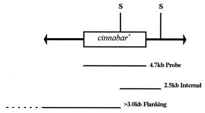
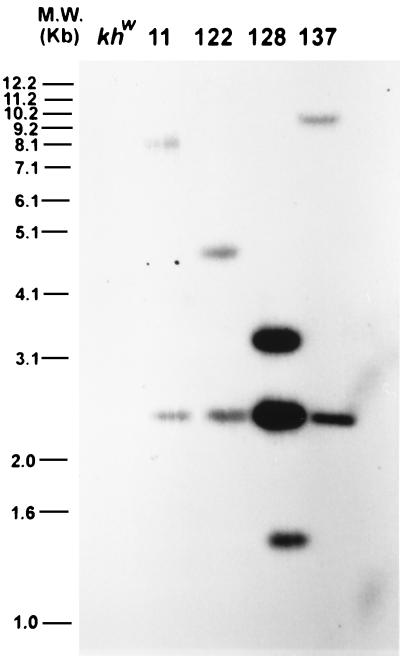
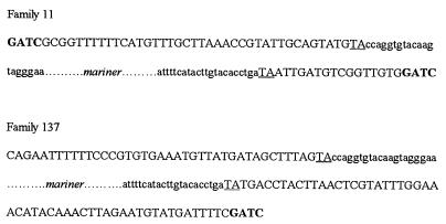
The mariner transposable element is capable of interplasmid transposition in the embryonic soma of the yellow fever mosquito, Aedes aegypti. To determine if this demonstrated mobility could be utilized to genetically transform the mosquito, a modified mariner element marked with a wild-type allele of the Drosophila melanogaster cinnabar gene was microinjected into embryos of a kynurenine hydroxylase-deficient, white-eyed recipient strain. Three of 69 fertile male founders resulting from the microinjected embryos produced families with colored-eyed progeny individuals, a transformation rate of 4%. The transgene-mediated complementation of eye color was observed to segregate in a Mendelian manner, although one insertion segregates with the recessive allele (female-determining) of the sex-determining locus, and a separate insertion is homozygous lethal. Molecular analysis of selected transformed families demonstrated that a single complete copy of the construct had integrated independently in each case and that it had done so in a transposase-mediated manner. The availability of a mariner transformation system greatly enhances our ability to study and manipulate this important vector species.


Figures

Figure 1
Figure 1
Schematic diagram of the mobile portion of the pM[cn] construct. The mariner inverted terminal repeats are represented as arrows flanking the 4.7-kb genomic DNA fragment from D. melanogaster that includes a copy of the cn+ gene. The relative positions of the SacI restriction endonuclease-cleavage sites are shown (S). Listed below are the relative extents and sizes of the fragment hybridized with the genomic DNA and the expected hybridizing genomic fragments from the transformed families.
Figure 2
Figure 2
Southern blot analysis of G2 genomic DNA from the transformed families, digested with SacI and hybridized with the 4.7-kb cn+ gene fragment. The predicted 2.5-kb internal fragment is observed in each case, as is a flanking fragment of greater than 3.0 kb, representing a single complete insertion event. Family 128 contains an additional 1.4-kb hybridizing fragment.
Figure 3
Figure 3
Primary DNA sequence of the junctions between the mariner inverted terminal repeats and the Ae. aegypti genomic DNA. The mariner sequence is shown in lowercase letters, the genomic Sau3AI sites are shown in bold (the complete left hand sequence from family 137 is not shown because the PCR product extends for several hundred base pairs), and the flanking TA residues are underlined.

References

    1. Collins F H, James A A. Sci Med. 1996;3:52–61.
    1. Jasinskiene, N., Coates, C. J., Benedict, M. Q., Cornel, A. J., Salazar Rafferty, C., James A. A. & Collins, F. H. (1998) Proc. Natl. Acad. Sci. USA, 3743–3747. - PMC - PubMed
    1. Sarkar A, Yardley K, Atkinson P W, James A A, O’Brochta D A. Insect Biochem Mol Biol. 1997;27:359–363. - PubMed
    1. Coates C J, Turney C L, Frommer M, O’Brochta D A, Atkinson P W. Mol Gen Genet. 1997;253:728–733. - PubMed
    1. Medhora M, Maruyama K, Hartl D L. Genetics. 1991;128:311–318. - PMC - PubMed

Publication types

LinkOut - more resources