. 1998 Mar 31;95(7):3758-63.
doi: 10.1073/pnas.95.7.3758.

Overlapping roles and asymmetrical cross-regulation of the USF proteins in mice

Affiliations

Overlapping roles and asymmetrical cross-regulation of the USF proteins in mice

M Sirito et al. Proc Natl Acad Sci U S A. .

Abstract

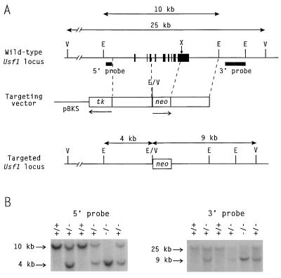
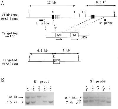
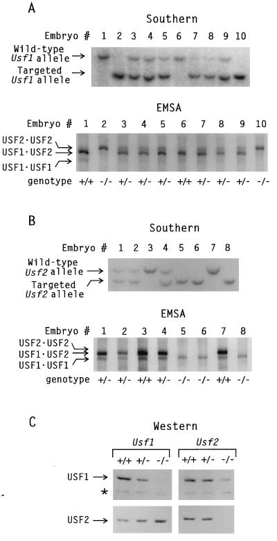
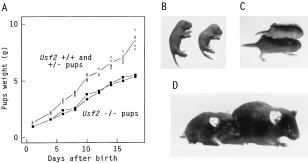
USF1 and USF2 are ubiquitously expressed transcription factors implicated as antagonists of the c-Myc protooncoprotein in the control of cellular proliferation. To determine the biological role of the USF proteins, mutant mice were generated by homologous recombination in embryonic stem cells. USF1-null mice were viable and fertile, with only slight behavioral abnormalities. However, these mice contained elevated levels of USF2, which may compensate for the absence of USF1. In contrast, USF2-null mice contained reduced levels of USF1 and displayed an obvious growth defect: they were 20-40% smaller at birth than their wild-type or heterozygous littermates and maintained a smaller size with proportionate features throughout postnatal development. Some of the USF-deficient mice, especially among the females, were prone to spontaneous epileptic seizures, suggesting that USF is important in normal brain function. Among the double mutants, an embryonic lethal phenotype was observed for mice that were homozygous for the Usf2 mutation and either heterozygous or homozygous for the Usf1 mutation, demonstrating that the USF proteins are essential in embryonic development.


Figures

Figure 1
Figure 1
Mutation of the Usf1 locus. (A) Targeting strategy. (Top) Structure of the wild-type Usf1 gene with solid boxes representing exons. The sizes of the restriction fragments detected by the indicated probes in wild-type DNA are shown above. E, EcoRI; V, EcoRV. (Middle) The gene-targeting vector. Open boxes, Usf1 homologous regions; neo, the PGK-neo expression cassette that introduces novel EcoRI and EcoRV restriction sites; tk, the MC1-tk expression cassette used for negative selection. The arrows beneath neo and tk indicate the direction of transcription of each cassette. (Bottom) Structure of the targeted locus, with the sizes of the restriction fragments detected by the Southern probes shown above. (B) Southern blot showing genotypes of newborn mice from a heterozygote mating. EcoRI-digested tail DNA was hybridized with the 3′ probe, and EcoRV-digested tail DNA was hybridized with the 5′ probe. The wild-type and mutant bands are shown in each case. Lanes: +/+, wild type; +/−, heterozygous mutant; −/−, homozygous mutant.
Figure 2
Figure 2
Mutation of the Usf2 locus. (A) Targeting strategy. Conventions are as indicated for Fig. 1. (B) Southern blot showing the genotypes of newborn mice. EcoRI-digested tail DNA was hybridized with the 5′ and 3′ probes. The wild-type and mutant bands are shown in each case. The asterisk indicates the migration of a band from a different locus that cross-reacted with the 3′ probe.
Figure 3
Figure 3
Expression of USF in mutant embryos. (A) A pregnancy resulting from the mating of Usf1(+/−) mice was interrupted at day 13.5 postconception, and MEF cell lines were established from each embryo. Genomic DNA was prepared from each cell line and used to establish the genotype of each embryo by Southern blotting. Mini nuclear extracts were prepared from each cell line and analyzed for USF DNA-binding species by EMSA, using a radiolabeled DNA fragment containing a USF-specific binding site under conditions that allow maximum resolution of the different USF dimers. Migration of protein–DNA complexes containing USF1 and USF2 polypeptides is indicated at left. (B) Same as in A but with embryos produced by Usf2(+/−) mice. (C) Nuclear extracts from MEFs of the indicated genotypes were analyzed by Western blotting using successively USF2- and USF1-specific antibodies. The asterisk indicates the migration of a USF-unrelated protein that interacted with the USF1 antibodies and represents an internal marker for equal protein loading in the different lanes.
Figure 4
Figure 4
Growth defect in Usf2(−/−) mutant mice. (A) All pups in one litter produced by a Usf2 heterozygous breeding pair were weighed at various times of postnatal development. Subsequent genotyping by tail DNA analysis revealed that the two smaller animals were homozygous mutants and that the wild-type and heterozygous pups all displayed similar weights. (B–D) Photographs of Usf2(−/−) mutants next to one of their siblings at birth (B) and at 6 days (C) or 1 month of age (D). Note the smaller size and proportionate body features of the mutants and the characteristic kinky tail of the newborn USF2-null pup.

References

    1. Sawadogo M, Roeder R G. Cell. 1985;43:165–175. - PubMed
    1. Carther R W, Chodosh L A, Sharp P A. Cell. 1985;43:439–448. - PubMed
    1. Miyamoto N G, Moncollin V, Egly J M, Chambon P. EMBO J. 1985;4:3563–3570. - PMC - PubMed
    1. Sawadogo M, Van Dyke M W, Gregor P D, Roeder R G. J Biol Chem. 1988;263:11985–11993. - PubMed
    1. Gregor P D, Sawadogo M, Roeder R G. Genes Dev. 1990;4:1730–1740. - PubMed

Publication types

LinkOut - more resources