. 1998 Mar 31;95(7):3792-7.
doi: 10.1073/pnas.95.7.3792.

NF-kappaB-inducing kinase activates IKK-alpha by phosphorylation of Ser-176

Affiliations

NF-kappaB-inducing kinase activates IKK-alpha by phosphorylation of Ser-176

L Ling et al. Proc Natl Acad Sci U S A. .

Abstract

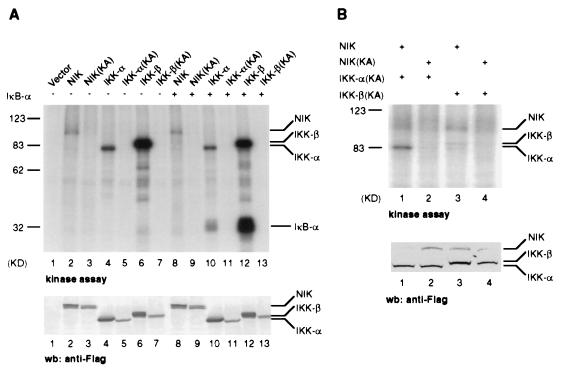
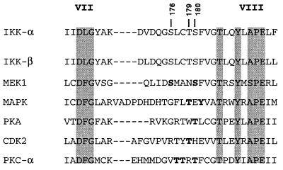
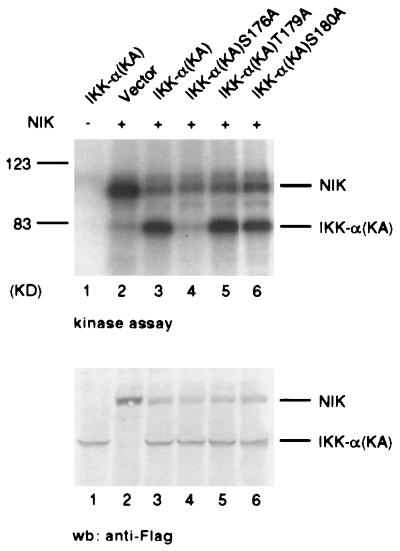
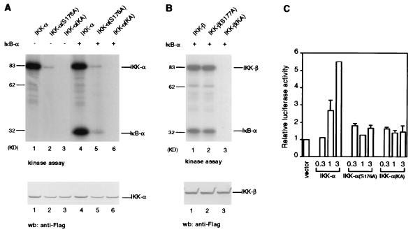
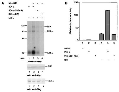
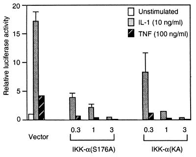
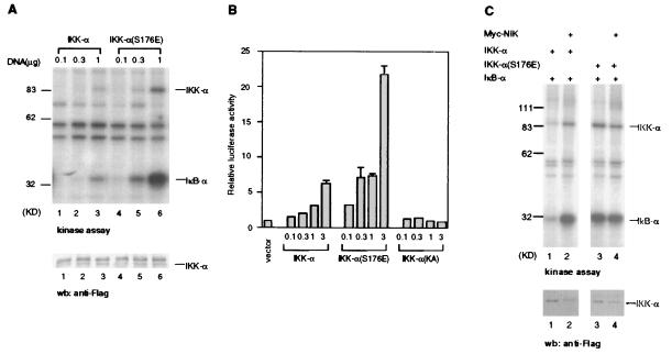
Activation of the transcription factor NF-kappaB by inflammatory cytokines involves the successive action of NF-kappaB-inducing kinase (NIK) and two IkappaB kinases, IKK-alpha and IKK-beta. Here we show that NIK preferentially phosphorylates IKK-alpha over IKK-beta, leading to the activation of IKK-alpha kinase activity. This phosphorylation of IKK-alpha occurs specifically on Ser-176 in the activation loop between kinase subdomains VII and VIII. A mutant form of IKK-alpha containing alanine at residue 176 cannot be phosphorylated or activated by NIK and acts as a dominant negative inhibitor of interleukin 1- and tumor necrosis factor-induced NF-kappaB activation. Conversely, a mutant form of IKK-alpha containing glutamic acid at residue 176 is constitutively active. Thus, the phosphorylation of IKK-alpha on Ser-176 by NIK may be required for cytokine-mediated NF-kappaB activation.


Figures

Figure 1
Figure 1
In vitro phosphorylation of IKK-α by NIK. (A) Autophosphorylation and phosphorylation of IκB-α by various kinases. 293 cells were transiently transfected with expression plasmids encoding FLAG epitope-tagged wild-type or KA mutants of NIK, IKK-α and IKK-β. Thirty-six hours after transfection, extracts were immunoprecipitated with anti-FLAG mAb affinity resin and FLAG-tagged proteins were purified as described in Materials and Methods. Purified proteins were incubated with [γ-32P]ATP in the presence or absence or bacterially synthesized protein IκB-α (amino acids 1–250), resolved by SDS/PAGE, and analyzed by autoradiography. The amounts of proteins used in the reactions were determined by immunoblotting (wb) with anti-FLAG polyclonal antibodies (Lower). The positions of IKK-α, IKK-β, and NIK are indicated. (B) Phosphorylation of IKK-α(KA) and IKK-β(KA) by NIK. 293 cells were transiently transfected with expression plasmids encoding FLAG epitope-tagged wild-type NIK, IKK-α(KA), or IKK-β(KA). Purified proteins were incubated with [γ-32P]ATP, resolved by SDS/PAGE, and analyzed by autoradiography. The amounts of proteins used in the reactions were determined by immunoblotting (wb) with anti-FLAG polyclonal antibodies (Lower). The positions of IKK-α, IKK-β, and NIK are indicated.
Figure 2
Figure 2
Alignment of IKK-α amino acid sequences with several kinases in the activation loop region. The D(F/L)G and (A/S)PE residues that are characteristic of kinase subdomains VII and VIII are shaded. The conserved threonine and tyrosine residues in the TXXY motif adjacent to subdomain VIII are also shaded. The activating phosphorylation sites in MEK1 (25, 26), MAPK (30), PKA (31), CDK2 (32), and PKC-α (33) are shown in boldface. The position of the serine and threonines residues of IKK-α are indicated. The sequence of the activation loop of IKK-β is also included.
Figure 3
Figure 3
Ser-176 in the activation loop of IKK-α is a major site of phosphorylation by NIK. Individual serine and threonine residues in the activation loop of IKK-α kinase domain were mutated to alanine. Each IKK-α mutant protein also contained the KA mutation in the ATP-binding site to prevent autophosphorylation. 293 cells were transiently transfected with expression plasmids encoding the indicated FLAG epitope-tagged proteins. Thirty-six hours after transfection, immunopurified proteins were incubated with [γ-32P]ATP, resolved by SDS/PAGE, and analyzed by autoradiography. The amount of protein used in each reaction was determined by immunoblotting (wb) with anti-FLAG polyclonal antibodies (Lower).
Figure 4
Figure 4
IKK-α(S176A) has reduced kinase activity and NF-κB activation. (A) IKK-α(S176A) has reduced kinase activity. 293 cells were transiently transfected with the indicated epitope-tagged expression vectors. Thirty-six hours after transfection, IKK-α proteins were immunopurified with anti-FLAG mAb affinity resin and used in in vitro kinase reactions with IκB-α and [γ-32P]ATP. (Lower) The protein expression in each lane is shown. (B) IKK-β(S177A) has similar kinase activity as IKK-β. 293 cells were transiently transfected with the indicated epitope-tagged expression vectors. Thirty-six hours after transfection, IKK-β proteins were immunopurified with anti-FLAG mAb affinity resin and used in in vitro kinase reactions with IκB-α and [γ-32P]ATP. (Lower) The protein expression in each lane is shown. (C) IKK-α(S176A) is defective in NF-κB activation. HeLa cells were transiently cotransfected with an E-selectin-luciferase reporter gene plasmid and vector control or IKK-α expression vector as indicated. Twenty-four hours after transfection, luciferase activities were determined and normalized on the basis of β-gal expression. The values shown are averages (±SEM) of duplicate samples for one representative experiment.
Figure 5
Figure 5
Loss of IKK-α(S176A) activation by NIK. (A) Loss of IKK-α(S176A) activation by NIK in kinase assay. 293 cells were transiently transfected with expression plasmids for FLAG epitope-tagged IKK-α or IKK-α(S176A) and Myc-epitope-tagged NIK. IKK-α proteins (and coprecipitating Myc-NIK proteins) were purified with anti-FLAG antibodies, and in vitro phosphorylation reactions were carried out by using bacterially expressed IκB-α and [γ-32P]ATP. The amounts of protein used were determined by immunoblotting with anti-Myc polyclonal antibodies (Middle), and with anti-FLAG polyclonal antibodies (Bottom). (B) Loss of IKK-α(S176A) activation by NIK in an NF-κB reporter gene assay. 293 cells were transiently cotransfected with an E-selectin-luciferase reporter gene plasmid and vector control or IKK-α and NIK expression vectors as indicated. Thirty to 36 hr after transfection, luciferase activities were determined and normalized on the basis of β-gal expression. The values shown are averages (±SEM) of duplicate samples for one representative experiment.
Figure 6
Figure 6
IKK-α(S176A) is a dominant negative inhibitor of IL-1 and TNF-induced NF-κB activation. 293/IL-1RI cells were transiently cotransfected with an E-selectin-luciferase reporter gene plasmid and vector control or IKK-α(S176A) expression vector as indicated. Twenty-four hours after transfection, cells were either left untreated, or stimulated for 6 hr with IL-1 (10 ng/ml) or TNF (100 ng/ml) prior to harvest. Luciferase activities were determined and normalized on the basis of β-gal expression. The values shown are averages (±SEM) of duplicate samples for one representative experiment.
Figure 7
Figure 7
IKK-α(S176E) is constitutively active. (A) IKK-α(S176E) has significantly greater activity than IKK-α in kinase assay. HeLa cells were transiently transfected with expression plasmids for FLAG epitope-tagged IKK-α or IKK-α(S176E) at different doses. Thirty hours later, IKK-α proteins were purified with anti-FLAG antibodies, and in vitro phosphorylation reactions were carried out with bacterially expressed IκB-α and [γ-32P]ATP. The amounts of protein used were determined by immunoblotting with anti-FLAG antibodies (as shown in the lower panel). (B) IKK-α(S176E) has significantly greater activity than IKK-α in an NF-κB reporter gene assay. HeLa cells were transiently cotransfected with an E-selectin-luciferase reporter gene plasmid and vector control or IKK-α expression vector as indicated. Twenty-four hours after transfection, luciferase activities were determined and normalized on the basis of β-gal expression. The values shown are averages (±SEM) of duplicate samples for one representative experiment. (C) IKK-α(S176E) activity is independent of NIK activation. HeLa cells were transiently transfected with expression plasmids for FLAG epitope-tagged IKK-α or IKK-α(S176E) and Myc-epitope-tagged NIK. IKK-α proteins were purified with anti-FLAG antibodies, and in vitro phosphorylation reactions were carried out by using bacterially expressed IκB-α and [γ-32P]ATP. The amounts of IKK-α protein used were determined by immunoblotting with anti-FLAG antibodies (Lower).

References

    1. Baeuerle P A, Henkel T. Annu Rev Immunol. 1994;12:141–179. - PubMed
    1. Baeuerle P A, Baltimore D. Cell. 1996;87:13–20. - PubMed
    1. Verma I M, Stevenson J K, Schwarz E M, Van Antwerp D, Miyamoto S. Genes Dev. 1995;9:2723–2735. - PubMed
    1. Traenckner E B, Pahl H L, Henkel T, Schmidt K N, Wilk S, Baeuerle P A. EMBO J. 1995;14:2876–2883. - PMC - PubMed
    1. Thanos D, Maniatis T. Cell. 1995;80:529–532. - PubMed