. 1998 Mar 31;95(7):3839-44.
doi: 10.1073/pnas.95.7.3839.

A conserved sequence block in murine and human T cell receptor (TCR) Jalpha region is a composite element that enhances TCR alpha enhancer activity and binds multiple nuclear factors

Affiliations

A conserved sequence block in murine and human T cell receptor (TCR) Jalpha region is a composite element that enhances TCR alpha enhancer activity and binds multiple nuclear factors

C L Kuo et al. Proc Natl Acad Sci U S A. .

Abstract

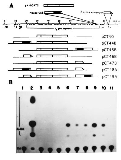
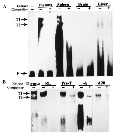
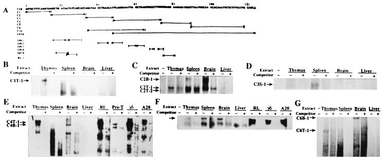
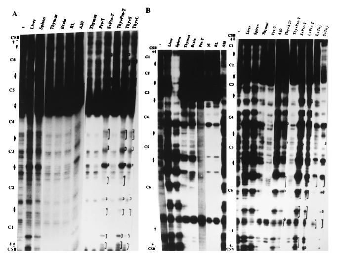
A conserved sequence block (CSB) located in a noncoding region of the mouse and human TCR alpha/delta loci, showing six differences over 125 nucleotide positions (95% similar), was subjected to detailed analyses in this study. Transient transfection results showed that the CSB-containing element in conjunction with the TCR alpha enhancer up-regulated the alpha enhancer activity, whereas no enhancer activity was detected when CSB alone was assayed. In vitro occupancy analyses of CSB by nuclear factors reveal the existence of an unexpectedly intricate network of CSB-protein and protein-protein interactions. Lymphoid-specific as well as T-lineage-specific nuclear factors are involved to differentially form CSB-bound complexes in extracts of various tissues and cell lines. Liver was shown to contain factor(s) sequestering thymic CSB-binding factors. Furthermore, the putative binding sites for transcription factors known to be important for lymphoid-lineage development are present in CSB and are targeted by nuclear factors. On the basis of these results, we propose that the CSB element may play a role in shaping the chromatin structure by which the accessibility of TCR alpha/delta loci to the recombinase complex and/or to the transcriptional apparatus can be controlled.


Figures

Figure 1
Figure 1
Effects of CSB-containing element on the expression of the CAT gene. (A) Schematic diagram of constructs used for the assays. pA10CAT2 is a fragment containing the partial SV40 promoter, the CAT gene, and the poly(A) site. It was inserted into pBluescript KS(−), and the resultant plasmid (pCT40) was used as the basic CAT vector. Mouse CSB is a CSB-containing element obtained by restriction enzyme digestion of mouse genomic DNA. The solid bar indicates the actual location of CSB in this fragment DNA. C alpha enhancer corresponds to a PvuII–BglII fragment containing the previously identified TCR α enhancer. The line below indicates the position of CSB and TCR α enhancer within the 95 kb of the mouse Cα/Cδ region. (B) Up-regulation of TCR α enhancer activity in αβ-expressing T cells. Constructs were electroporated into EL4 cells. The cells were harvested and samples were normalized with respect to total protein prior to performing the CAT assay. The acetylated chloramphenicol levels were measured qualitatively by autoradiography and quantitatively by a phosphoimager. The quantitative values are given in parenthesis below. Lane 1, assay blank (0); lane 2, E. coli CAT as a positive control (1,350); lane 3, baseline construct (pCT40) (16); lane 4, CSB in front of CAT gene (pCT44B) (12); lane 5, CSB in back of CAT gene (pCT45B) (0); lane 6, α enhancer in front (pCT46B) (176); lane 7, α enhancer in back (pCT47B) (74); lane 8, CSB in front, α enhancer in back (pCT48A) (90); lane 9, α enhancer in front, CSB in back (pCT49A) (333); lane 10, SV40 enhancer in front (138); and lane 11, mock transfection control (0). Chl indicates unacetylated [14C]chloramphenicol; Ac-Chl indicates acetylated [14C]chloramphenicol.
Figure 2
Figure 2
Binding of nuclear factors to the 125-bp CSB element. (A) Tissue-specific binding to CSB. 32P-labeled CSB was incubated with 5 μg of nuclear extracts prepared from adult mouse thymus, spleen, liver, or brain to perform EMSAs. No extract was added to the substrate in the first lane. Free probe (F) and CSB–factor complexes were resolved electrophoretically; the resulting autoradiogram is shown. The shifted complexes seen with the thymus extract are designated T1 and T2. Assays were done in the absence (−) or presence (+) of 50-fold excess of unlabeled CSB as specific competitor. (B) Cell type-specific binding to CSB. EMSA was performed as detailed for A except 32P-labeled CSB was incubated with nuclear extract prepared from various cultured lines of T (RL, a leukemia T cell line; Pre-T, pre-T cell line SCI.ET.29F; γδ, a TCR γδ-expressing hybridoma GP39) and B (A20, a B cell lymphoma) cells.
Figure 3
Figure 3
Fine mapping of nuclear factor recognition sites within CSB. (A) The sequence of the CSB element (125 bp in length) is shown at the top. Below that are arrowhead-lines representing different subsites of CSB, including subregions assayed for bindings of nuclear factors by EMSA. They are C1, C2, C3, C4, C5, C6, C34, and C456. The inverted repeats (denoted as INV1 to INV4), the putative GATA-binding site (denoted as GATA), and PU.1-binding site (denoted as PU.1) are also presented (see text). (B–G) Nuclear extracts prepared from various adult mouse tissue and/or a series of cultured cell lines were incubated with 32P-labeled C1 (B), C2 (C), C3 (D), C4 (E), C5 (F), or C6 (G) to perform EMSAs. Assays were done in the absence (−) or presence (+) of 50-fold excess of unlabeled C1 (B), C2 (C), C3 (D), C4 (E), C5 (F), or C6 (G) as competitor. Free probe and CSB subregion–factor complexes were resolved by electrophoresis. Only the relevant resulting autoradiograms are shown. C1T-1, the major C1-binding complex formed in thymus; C2B-1, the slowest migrating C2-binding complex formed in brain; C2T-1 and C2T-2, the two major C2-binding complexes formed in thymus; C3S-1, the C3-binding complex formed in spleen; C4T-1 and C4B-1, the unique C4-binding complexes formed in thymus and brain, respectively; and C6B-1 and C6T-1, the major unique C6-binding complexes formed in brain and thymus, respectively.
Figure 4
Figure 4
Comparison of footprinting patterns established on CSB by nuclear extracts of various mouse tissues and cultured cell lines. A CSB-containing fragment was analyzed by DNase I footprinting, as described in the text. The autoradiograms of DNase I digestion of the top strand and the bottom strand are shown in (A) and (B), respectively. Nuclear extracts from various tissues and cell lines, and mixed nuclear extracts of spleen and pre-T (S+Pre-T), thymus and pre-T (Thy+Pre-T), thymus and spleen (Thy+S), thymus and liver (Thy+L), thymus and A20 (Thy+A20), or liver and pre-T (L+Pre-T) were used in the assays. −, No nuclear extract. The regions corresponding to subregions of CSB, shown in Fig. 3A, are indicated on the side of each gel. The bracketed regions indicate the regions with footprint pattern changes in mixed nuclear extracts when compared with the pattern produced by thymus, pre-T, or Thy+Pre-T.

References

    1. Tonegawa S. Nature (London) 1983;302:575–581. - PubMed
    1. Kronenberg M, Siu G, Hood L, Shastri N. Annu Rev Immunol. 1986;4:529–591. - PubMed
    1. Raulet D H. Annu Rev Immunol. 1989;7:175–207. - PubMed
    1. Honjo T. Annu Rev Immunol. 1983;1:499–528. - PubMed
    1. Yancopoulos G, Blackwell H, Suh H, Hood L, Alt F W. Cell. 1986;44:251–259. - PubMed

Publication types

MeSH terms

Substances

LinkOut - more resources