. 1998 Mar 31;95(7):3862-6.
doi: 10.1073/pnas.95.7.3862.

An X chromosome gene regulates hematopoietic stem cell kinetics

Affiliations

An X chromosome gene regulates hematopoietic stem cell kinetics

J L Abkowitz et al. Proc Natl Acad Sci U S A. .

Abstract

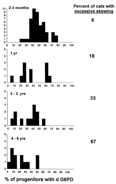
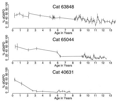
Females are natural mosaics for X chromosome-linked genes. As X chromosome inactivation occurs randomly, the ratio of parental phenotypes among blood cells is approximately 1:1. Recently, however, ratios of greater than 3:1 have been observed in 38-56% of women over age 60. This could result from a depletion of hematopoietic stem cells (HSCs) with aging (and the maintenance of hematopoiesis by a few residual clones) or from myelodysplasia (the dominance of a neoplastic clone). Each possibility has major implications for chemotherapy and for transplantation in elderly patients. We report similar findings in longitudinal studies of female Safari cats and demonstrate that the excessive skewing that develops with aging results from a third mechanism that has no pathologic consequence, hemizygous selection. We show that there is a competitive advantage for all HSCs with a specific X chromosome phenotype and, thus, demonstrate that an X chromosome gene (or genes) regulates HSC replication, differentiation, and/or survival.


Figures

Figure 1
Figure 1
Distribution of G6PD phenotype among progenitor cells from female Safari cats at various ages. The data at 2–3 months represent studies in 48 female Safari cats. Because most animals were entered into studies of retroviral physiology or hematopoiesis after baseline evaluations, the numbers of normal (unmanipulated) animals in the subsequent studies are smaller. Excessive skewing is defined as a distribution of G6PD phenotype in which <25% of progenitors contained d G6PD or >75% of progenitors contained d G6PD. The percent of animals with excessive skewing increased with age. This skewing was toward the G G6PD phenotype (resulting in a smaller percentage of progenitors with d G6PD). A significant change from a binomial distribution is seen in the 2- to 3-year and 4- to 6-year data when assessed by Q–Q plots (12) (analysis not shown). The X chromosome inactivation pattern observed at 2–3 months is similar to that reported in human studies (13, 14). By using the approach of these studies, the data can be further analyzed. If X chromosome inactivation took place after k divisions, 2k cells would be present at that time. Given binomial variability, the distribution would have a standard deviation of 2−(1 + k/2) × 100%, because the sample size is 2k and the probability of d G6PD is 1/2. When we equate this description of a binomial distribution with the observed data and solve for k, we estimate that k equals 4. Thus, approximately 16 cells were present at the time of X chromosome inactivation, or alternatively 16 cells gave rise to the hematopoietic system during development, similar to calculations in the referenced human studies.
Figure 2
Figure 2
Longitudinal studies of progenitor cells from three representative female Safari cats. In cat 63848, a slow change toward a dominance of progenitor cells with a G G6PD phenotype was seen. Significant deviation from the mean value was first observed at age 8.4 years, when the data are analyzed by sequential χ2 analyses. In cat 65044, a significant change in the percent of progenitors with d G6PD was first noted at age 6.3 years, and a change in the phenotype of cells from cat 40631 was evident by year 2.7. Although a similar drift occurred among red cells and granulocytes (data not shown), the G6PD phenotype of T cells remained unchanged and specifically was 60% (year 13) in cat 63848, 60% (year 12) in cat 65044, and 60% (year 7) in cat 40631.

References

    1. Orlic D, Bodine D M. Blood. 1994;84:3991–3994. - PubMed
    1. Abkowitz J L, Catlin S N, Guttorp P. Nat Med. 1996;2:190–197. - PubMed
    1. Wang J C Y, Doedens M, Dick J E. Blood. 1997;89:3919–3924. - PubMed
    1. Busque L, Mio R, Mattioli J, Brais E, Blais N, Lalonde Y, Maragh M, Gilliland D G. Blood. 1996;88:59–65. - PubMed
    1. Gale R E, Fielding A K, Harrison C N, Linch D C. Br J Haematol. 1997;98:512–519. - PubMed

Publication types