

Deregulated expression of TCL1 causes T cell leukemia in mice
- PMID: 9520462
- PMCID: PMC19932
- DOI: 10.1073/pnas.95.7.3885
Deregulated expression of TCL1 causes T cell leukemia in mice
Abstract
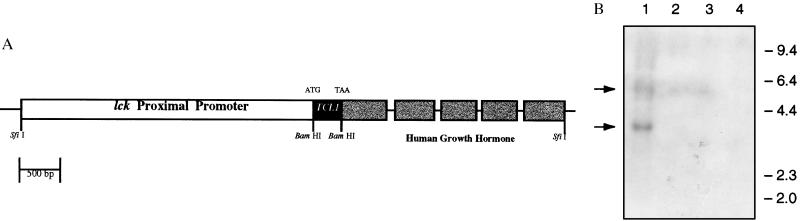
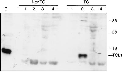
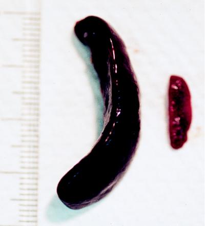
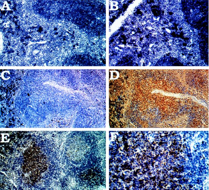
The TCL1 oncogene on human chromosome 14q32.1 is involved in the development of T cell leukemia in humans. These leukemias are classified either as T prolymphocytic leukemias, which occur very late in life, or as T chronic lymphocytic leukemias, which often arise in patients with ataxia telangiectasia (AT) at a young age. The TCL1 oncogene is activated in these leukemias by juxtaposition to the alpha or beta locus of the T cell receptor, caused by chromosomal translocations t(14:14)(q11:q32), t(7:14)(q35:q32), or by inversions inv(14)(q11:q32). To show that transcriptional alteration of TCL1 is causally involved in the generation of T cell neoplasia we have generated transgenic mice that carry the TCL1 gene under the transcriptional control of the p56(lck) promoter element. The lck-TCL1 transgenic mice developed mature T cell leukemias after a long latency period. Younger mice presented preleukemic T cell expansions expressing TCL1, and leukemias developed only at an older age. The phenotype of the murine leukemias is CD4-CD8+, in contrast to human leukemias, which are predominantly CD4+CD8-. These studies demonstrate that transcriptional activation of the TCL1 protooncogene can cause malignant transformation of T lymphocytes, indicating the role of TCL1 in the initiation of malignant transformation in T prolymphocytic leukemias and T chronic lymphocytic leukemias.
Figures
Similar articles
-
Human chronic lymphocytic leukemia modeled in mouse by targeted TCL1 expression.Proc Natl Acad Sci U S A. 2002 May 14;99(10):6955-60. doi: 10.1073/pnas.102181599. Proc Natl Acad Sci U S A. 2002. PMID: 12011454 Free PMC article.
-
TCL1 oncogene activation in preleukemic T cells from a case of ataxia-telangiectasia.Blood. 1995 Sep 15;86(6):2358-64. Blood. 1995. PMID: 7662982
-
Role of TCL1 and ALL1 in human leukemias and development.Cancer Res. 1999 Apr 1;59(7 Suppl):1778s-1783s. Cancer Res. 1999. PMID: 10197596 Review.
-
TCL1 is overexpressed in patients affected by adult T-cell leukemias.Cancer Res. 1997 Dec 15;57(24):5452-6. Cancer Res. 1997. PMID: 9407948
-
The role of TCL1 in human T-cell leukemia.Oncogene. 2001 Sep 10;20(40):5638-43. doi: 10.1038/sj.onc.1204596. Oncogene. 2001. PMID: 11607815 Review.
Cited by
-
Germline risk of clonal haematopoiesis.Nat Rev Genet. 2021 Sep;22(9):603-617. doi: 10.1038/s41576-021-00356-6. Epub 2021 May 13. Nat Rev Genet. 2021. PMID: 33986496 Free PMC article. Review.
-
TCL1: a shared tumor-associated antigen for immunotherapy against B-cell lymphomas.Blood. 2012 Aug 23;120(8):1613-23. doi: 10.1182/blood-2011-09-382838. Epub 2012 May 29. Blood. 2012. PMID: 22645177 Free PMC article.
-
T-cell prolymphocytic leukemia.Proc (Bayl Univ Med Cent). 2013 Jan;26(1):19-21. doi: 10.1080/08998280.2013.11928902. Proc (Bayl Univ Med Cent). 2013. PMID: 23382603 Free PMC article.
-
Protooncogene TCL1b functions as an Akt kinase co-activator that exhibits oncogenic potency in vivo.Oncogenesis. 2013 Sep 16;2(9):e70. doi: 10.1038/oncsis.2013.30. Oncogenesis. 2013. PMID: 24042734 Free PMC article.
-
Expression of TCL-1 as a potential prognostic factor for treatment outcome in B-cell chronic lymphocytic leukemia.Leuk Res. 2007 Dec;31(12):1737-40. doi: 10.1016/j.leukres.2007.05.020. Epub 2007 Jul 19. Leuk Res. 2007. PMID: 17659340 Free PMC article. Clinical Trial.
References
Publication types
MeSH terms
Substances
Grants and funding
LinkOut - more resources
Full Text Sources
Other Literature Sources
Molecular Biology Databases
Research Materials
Miscellaneous
