. 1998 Mar 31;95(7):3908-13.
doi: 10.1073/pnas.95.7.3908.

Engraftment and migration of human bone marrow stromal cells implanted in the brains of albino rats--similarities to astrocyte grafts

Affiliations

Engraftment and migration of human bone marrow stromal cells implanted in the brains of albino rats--similarities to astrocyte grafts

S A Azizi et al. Proc Natl Acad Sci U S A. .

Abstract

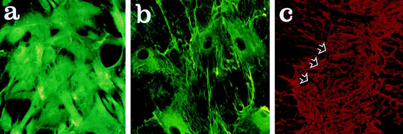
Neurotransplantation has been used to explore the development of the central nervous system and for repair of diseased tissue in conditions such as Parkinson's disease. Here, we examine the effects of direct injection into rat brain of human marrow stromal cells (MSCs), a subset of cells from bone marrow that include stem-like precursors for nonhematopoietic tissues. Human MSCs isolated by their adherence to plastic were infused into the corpus striatum. Five to 72 days later, brain sections were examined for the presence of the donor cells. About 20% of the infused cells had engrafted. There was no evidence of an inflammatory response or rejection. The cells had migrated from the injection site along known pathways for migration of neural stem cells to successive layers of the brain. After infusion into the brain, the human MSCs lost their immunoreactivity to antibodies for collagen I. Initially, the human cells continued to stain with antibodies to fibronectin but the region of staining with fibronectin was significantly decreased at 30 and 72 days. The results suggest that MSCs may be useful vehicles for autotransplantation in both cell and gene therapy for a variety of diseases of the central nervous system.


Figures

Figure 1
Figure 1
Effect of PDGF-AA on growth of human MSCs. Values are means ± SD (n = 3). ⧫, control; ▴, 1 ng/ml PDGF; □, 5 ng/ml PDGF; ▪, 10 ng/ml PDGF.
Figure 2
Figure 2
Photomicrographs that demonstrate some of the morphological characteristics of MSCs and astrocytes in culture. a and b demonstrate two types of human MSCs, flat and elongated. c demonstrates the similar morphology of rat MSCs. d shows fluorescent labeling of nuclei of human MSCs immediately before implantation. e and f demonstrate indirect immunofluorescent staining of astrocytes with antibodies against vimentin and glial fibrillary acidic protein, respectively. (Magnification: a, b and c, ×20; d, ×10; e and f, ×40.)
Figure 3
Figure 3
Photomicrographs that demonstrate sites of implantation of bone MSCs in the striatum of adult rats. (a) Site of implantation of human MSCs after 14 days. Arrows indicate needle track. Hemotoxylin and eosin stain. (b) Adjacent section to a stained with antibodies to HLA-ABC. (c) Adjacent section to a examined for nuclear fluorescence of human MSCs. Arrows indicate needle track. (d) Site of implantation of human MSCs after 72 days. Section examined for nuclear fluorescence. (e) Same as d 30 days after implantation. (f) Site of implantation of rat MSCs 14 days after implantation. Examined for nuclear fluorescence. (Magnification: a and b, ×10; cf, ×4.)
Figure 4
Figure 4
Line drawings of rat forebrain demonstrate the extent of migration of human MSCs and astrocytes after implantation in the striatum of the rat at the level of bregma. The drawing is a composite from brains examined at 4, 14, 30, and 72 days after the cell infusions. The pattern of implantation and migration of human MSCs is similar to that of rat astrocytes. The outlying cells, located in the temporal cortex and areas of corpus callosum farthest from the injection sites, were first to disappear. •, clusters of human MSCs; ○, clusters of rat astrocytes.
Figure 5
Figure 5
Photomicrographs that demonstrate staining with antibodies to collagen I and fibronectin. (a) Human MSCs in culture stained for collagen I. (b) Same stained for fibronectin. (c) Section of rat brain stained for fibronectin 5 days after implantation of human MSCs. Arrows indicate deposits of fibronectin. (Magnification: a and b, ×20; c, ×4.)

References

    1. McKay R. Science. 1997;276:66–71. - PubMed
    1. Bjorklund A. Nature (London) 1993;362:414–415. - PubMed
    1. Olson L. Nat Med. 1997;3:1329–1335. - PubMed
    1. Spencer D D, Robbins R J, Naftolin F, Marek K L, Vollmer T, Leranth C, Roth R H, Price L H, Gjedde A, Bunney B S, et al. N Engl J Med. 1992;327:1541–1548. - PubMed
    1. Freed C R, Breeze R E, Rosenberg N L, Schneck S A, Kriek E, Oi J X, Lone T, Zhang U B, Snyder J A, Wells T H, et al. N Engl J Med. 1992;327:1549–1555. - PubMed

Publication types

LinkOut - more resources