. 1998 Mar 31;95(7):3914-9.
doi: 10.1073/pnas.95.7.3914.

Budesonide epimer R or dexamethasone selectively inhibit platelet-activating factor-induced or interleukin 1beta-induced DNA binding activity of cis-acting transcription factors and cyclooxygenase-2 gene expression in human epidermal keratinocytes

Affiliations

Budesonide epimer R or dexamethasone selectively inhibit platelet-activating factor-induced or interleukin 1beta-induced DNA binding activity of cis-acting transcription factors and cyclooxygenase-2 gene expression in human epidermal keratinocytes

W J Lukiw et al. Proc Natl Acad Sci U S A. .

Abstract

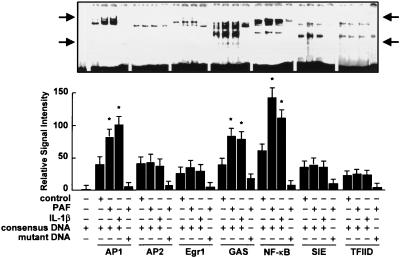
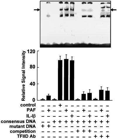
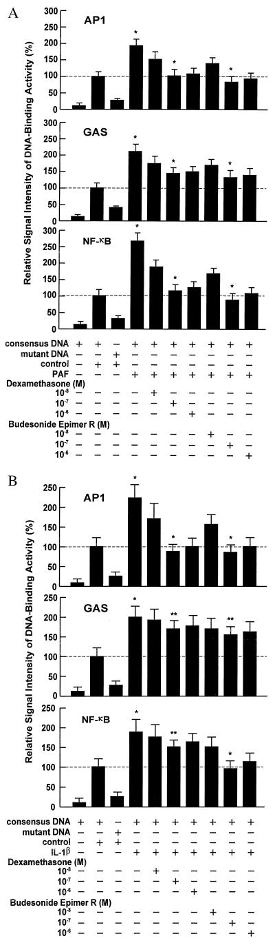
To further understand the molecular mechanism of glucocorticoid action on gene expression, DNA-binding activities of the cis-acting transcription factors activator protein 1 (AP1), AP2, Egr1 (zif268), NF-kappaB, the signal transducers and activators of transcription proteins gamma interferon activation site (GAS), Sis-inducible element, and the TATA binding protein transcription factor II D (TFIID) were examined in human epidermal keratinocytes. The cytokine interleukin 1beta (IL-1beta) and platelet-activating factor (PAF), both potent mediators of inflammation, were used as triggers for gene expression. Budesonide epimer R (BUDeR) and dexamethasone (DEX) were studied as potential antagonists. BUDeR or DEX before IL-1beta- or PAF-mediated gene induction elicited strong inhibition of AP1-, GAS-, and in particular NF-kappaB-DNA binding (P < 0.001, ANOVA). Only small effects were noted on AP2, Egr1 (zif268), and Sis-inducible element-DNA binding (P > 0.05). No significant effect was noted on the basal transcription factor TFIID recognition of TATA-containing core promoter sequences (P > 0.68). To test the hypothesis that changing cis-acting transcription factor binding activity may be involved in inflammatory-response related gene transcription, RNA message abundance for human cyclooxygenase (COX)-1 and -2 (E.C.1.14.99.1) was assessed in parallel by using reverse transcription-PCR. Although the COX-1 gene was found to be expressed at constitutively low levels, the TATA-containing COX-2 gene, which contains AP1-like, GAS, and NF-kappaB DNA-binding sites in its immediate promoter, was found to be strongly induced by IL-1beta or PAF (P < 0.001). BUDeR and DEX both suppressed COX-2 RNA message generation; however, no correlation was associated with TFIID-DNA binding. These results suggest that on stimulation by mediators of inflammation, although the basal transcription machinery remains intact, modulation of cis-activating transcription factor AP1, GAS, and NF-kappaB-DNA binding by the glucocorticoids BUDeR and DEX play important regulatory roles in the extent of specific promoter activation and hence the expression of key genes involved in the inflammatory response.


Figures

Figure 1
Figure 1
PAF or IL-1β differentially enhance DNA binding activities of cis-acting transcription factors. EMSA using NPXTs from HEK cells reacted with target DNA oligonucleotide consensus or with mutant sequences for the transcription factors AP1, AP2, Egr1 (zif268), GAS, NF-κB, SIE, and TFIID described in Table 1. Leftmost lane represents the migration of a typical free 32P end-labeled oligonucleotide with no NPXT. Relative gel shift signal quantitation in the bar graph was for gel-shifted species unique to that transcription factor-DNA binding consensus sequence, minus the signal contributed by the mutant DNA, and located in between the two sets of arrows. NF-κB quantitation includes only the upper (p50-p65) activator complexes whose identity was confirmed by using p50 and p65 antibodies and gel supershift assay (data not shown). Gel is overexposed to show the relative signal intensities among each of the shifted species. Exposure time = 12 hr. n = 4; mean ± SD; significance of induced factors over control, ∗, P < 0.001; significance of AP2, Egr1, and SIE over control P > 0.05; significance of TFIID over control, P > 0.68, ANOVA.
Figure 2
Figure 2
PAF or IL-1β do not modify TFIID-DNA binding. EMSA using NPXTs derived from HEK cells and the mutant and consensus sequence for the basal transcription factor TFIID. Arrow depicts unique TFIID-shifted complex (34, 45) used for signal quantitation in the bar graph shown below. A 60-fold excess of unlabeled TFIID consensus sequence (“cold” competition assay) almost completely eliminated TFIID gel shift. Rabbit polyclonal antibody specific for human TFIID was used in the gel supershift assay. Exposure time = 36 hr. n = 4; mean ± SD; significance of PAF- or IL-1β-induced TFIID factor over control, P > 0.68.
Figure 3
Figure 3
Dose-dependent inhibition by BUDeR or DEX of DNA binding by cis-acting transcription factors AP1, GAS, and NF-κB in HEK cells activated by PAF (A) or IL-1β (B). Relative signal intensity of DNA binding activity as determined by EMSA using NPXTs derived from HEK cells and reacted with the target oligonucleotide consensus sequences for the transcription factors AP1, GAS, or NF-κB. Leftmost lanes represent the relative signal intensity of a typical free oligonucleotide with no NPXT. Dashed line represents HEK control NPXT and DNA consensus sequences for transcription factors AP1, GAS, or NF-κB (Table 1) equal to 100. Compared with controls, both PAF (A) and IL-1β (B) elicited a 1.9- to 2.6-fold increase in transcription factor-DNA binding for AP1, GAS, and NF-κB. (M) = molar. n = 4; mean ± SD; significance of induced factors over control: ∗, P < 0.001, ∗∗, P < 0.01, ANOVA. Levels of TFIID-DNA binding remained consistently unchanged (P ≥ 0.68, ANOVA; Figs. 1 and 2).
Figure 4
Figure 4
PAF or IL-1β, but not 1L-6, induces COX-2 gene expression in HEK cells. Dose-dependent inhibition of this induction by BUDeR or DEX. (A) Diagram of the human COX-2 promoter showing transcription factor DNA binding consensus sequences from −1,000 and +165 bp relative to the start of transcription (+1 bp). DNA binding consensus sequences are described in Tables 1 and 2. Note that the TFIID site doubles as a GAS binding site. (B) Representative gel showing COX-1 and COX-2 RNA message abundance after treatment with PAF (100 nM), IL-1β (10 ng/ml), IL-6 (10 ng/ml), or DEX and BUDeR (each at 10−7 M). (C) RNA message levels quantitated from B. n = 4; mean ± SD; significance of induced RNA levels over control, ∗, P < 0.001, ∗∗, P > 0.05, ANOVA. (D) Dose-dependent inhibition by BUDeR or DEX on PAF- or IL-1β-induced changes on COX-1 and COX-2 RNA message abundance. n = 4; mean ± SD; significance of GC suppression at 10−7 M compared with no treatment, ∗, P < 0.001, ANOVA.

References

    1. Barnes P J, Adcock I. Trends Pharmacol Sci. 1993;14:436–441. - PubMed
    1. Tsai M J, O’Malley B W. Annu Rev Biochem. 1994;63:451–486. - PubMed
    1. Bamberger C M, Schulte H M, Chrousos G P. Endocr Rev. 1996;17:245–261. - PubMed
    1. Wilckens T, De Rijk R. Immunol Today. 1997;18:418–424. - PubMed
    1. Kerppola T K, Luk D, Curran T. Mol Cell Biol. 1993;13:3782–3791. - PMC - PubMed

MeSH terms