. 1998 Mar 31;95(7):3931-6.
doi: 10.1073/pnas.95.7.3931.

Herpes simplex virus 1 induces and blocks apoptosis at multiple steps during infection and protects cells from exogenous inducers in a cell-type-dependent manner

Affiliations

Herpes simplex virus 1 induces and blocks apoptosis at multiple steps during infection and protects cells from exogenous inducers in a cell-type-dependent manner

V Galvan et al. Proc Natl Acad Sci U S A. .

Abstract

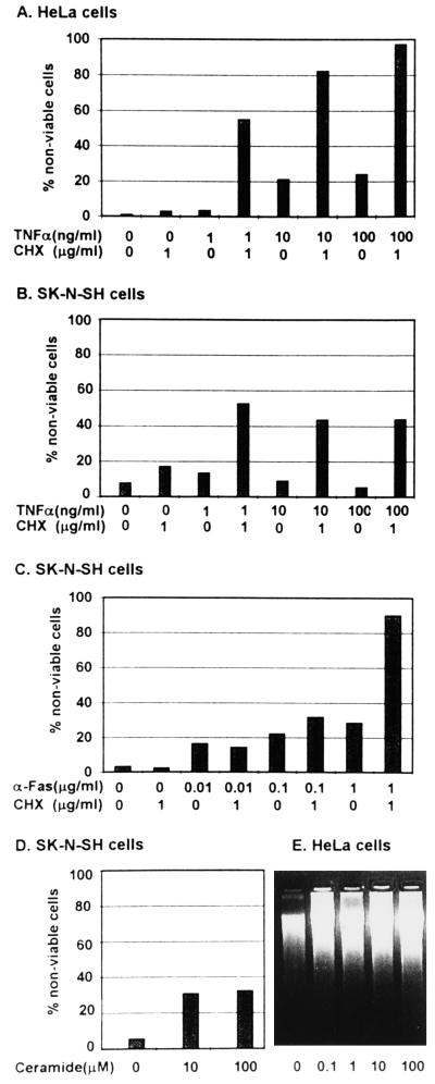
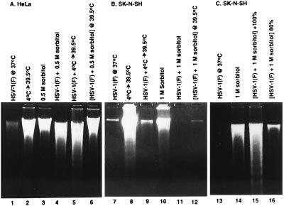
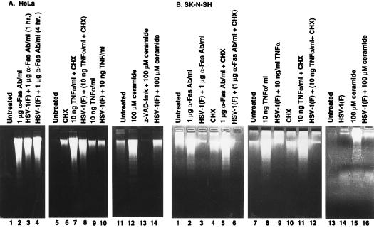
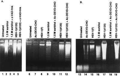
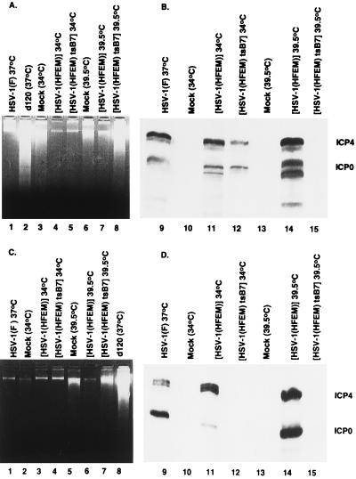
Several publications have attested to the ability of herpes simplex viruses to protect cells against apoptosis. We investigated the ability of the virus to protect cells in continuous cultivation from apoptosis induced by the virus itself, and by other known inducers such as exposure to the tumor necrosis factor alpha (TNFalpha), antibody to Fas, C2-ceramide, osmotic shock (sorbitol), and thermal shock. The salient features of the results were that the virus was able to protect cells against apoptosis by all of the agents tested, and that apoptosis induced by the virus was a very early event that did not require de novo expression of viral genes. However, these events were cell-type specific. Thus: (i) The cell lines tested exhibited fragmented chromosomal DNA following infection with a virus lacking functional alpha4 and US3 genes encoding the major regulatory protein and a viral protein kinase, respectively, but not by wild-type virus. (ii) Wild-type virus protected subconfluent SK-N-SH but not HeLa cells against induction of apoptosis by anti-Fas antibody, TNFalpha, C2-ceramide, and thermal shock. Confluent SK-N-SH cells were not protected from osmotic shock-induced apoptosis by wild-type infection. (iii) Wild-type virus protected SK-N-SH but not HeLa cells against induction of apoptosis by sorbitol, anti-Fas antibody, or TNFalpha and C2-ceramide. (iv) Mutant HSV-1(HFEM)tsB7 at the nonpermissive temperature infects cells but the DNA is not released from capsids, and therefore viral gene expression is restricted to the function of viral proteins introduced into the cell along with the capsid containing the viral DNA. HSV-1(HFEM)tsB7 induced apoptosis in Vero cells but not in SK-N-SH cells infected and maintained at 39.5 degrees C. (v) Tests of two caspase inhibitors showed that they blocked apoptosis induced by C2-ceramide and sorbitol, but were not able to block apoptosis induced by the virus lacking functional alpha4 and US3 genes. We conclude that HSV-1 triggers apoptosis at multiple metabolic checkpoints and in turn has evolved mechanisms to block apoptosis at each point and that some of the pathways of induction are shared with exogenous inducers tested in this study whereas others are not.


Figures

Figure 1
Figure 1
Induction of apoptosis by anti-Fas antibody, TNFα and C2-ceramide. (A–C) Cells were exposed to TNFα, or anti-Fas antibody alone or in the presence of CHX and assayed for viability. (D) SK-N-SH cells were exposed to C2-ceramide and tested for viability. (E) HeLa cells exposed to C2-ceramide and tested for degradation of DNA. The procedures were as described in Materials and Methods.
Figure 2
Figure 2
Photograph of agarose gels containing electrophoretically separated low molecular weight DNA fractions from mock-infected or HSV-1(F)-infected cells and stained with ethidium bromide. Subconfluent HeLa (A) or SK-N-SH cells (B) or subconfluent and confluent SK-N-SH cells (C) were mock-infected (lanes 2, 3, 8, 10 and 14) or infected with HSV-1(F) (lanes 1, 4–7, 9, 11–13, 15 and 16), treated by temperature shift (4°C → 39.5°C) or osmotic shock (0.5 M or 1 M sorbitol) and incubated for 5 hr (sorbitol-treated cells) or for 36 hr (other cultures) at 37°C unless otherwise indicated. 2 × 10⁶ cells per sample were collected, rinsed in PBS and lysed in a solution containing 10 mM Tris⋅HCl (pH 8.0), 10 mM EDTA and 0.5% Triton X-100 and centrifuged at 14,000 rpm for 30 min in an Eppendorf microcentrifuge to pellet chromosomal DNA. The supernatant fluids were digested with 0.1 mg/ml RNase A at 37°C for 1 hr and then with 1 mg/ml proteinase K at 50°C in the presence of 1% SDS, extracted with phenol and chloroform, precipitated in cold ethanol and subjected to electrophoresis in 2% agarose gels containing 5 μg/ml ethidium bromide. DNA was visualized by UV light transillumination and photographed with the Eagle Eye II (Stratagene).
Figure 3
Figure 3
Photograph of agarose gels containing electrophoretically separated low molecular weight DNA fractions from mock-infected or infected cells and stained with ethidium bromide. Subconfluent HeLa (A) or SK-N-SH cells (B) were mock-infected or infected with HSV-1(F) and treated by incubation in 1 μg/ml of anti-Fas IgM or 10 ng/ml TNFα both in the presence or the absence of 1 μg/ml CHX. Anti-Fas IgM or TNFα-treated HeLa cells were collected at 8 and 6 hr after treatment, respectively. The SK-N-SH cell samples were collected at 12 and 16 hr after treatment, respectively. Mock-infected and infected HeLa and SK-N-SH cells were treated with 100 μM C2-ceramide and collected at 6 hr after treatment.
Figure 4
Figure 4
(A) Photograph of agarose gels containing electrophoretically separated low molecular weight DNA fractions from mock-infected cells or cells infected with d120 mutant and stained with ethidium bromide. Subconfluent Vero (A) or HeLa cells (B) were either left untreated or treated with 50 μM z-VAD-fmk or 100 μM Ac-DEVD-CHO (Vero cells) or treated with 100 μM Ac-DEVD-CHO (HeLa cells) and then exposed to 1.5 M sorbitol or infected with 10 pfu of d120 mutant per cell and harvested at 5 hr after treatment and 24 hr after infection, respectively. Samples were processed as described in the legend to Fig. 2.
Figure 5
Figure 5
(A) Photograph of agarose gels containing electrophoretically separated, ethidium bromide stained low molecular weight DNA (A and C) or of immunoblots (B and D). Subconfluent Vero cells (A) or SK-N-SH cells (C) were mock-infected or infected with 10 pfu of HSV-1(F), d120, HSV-1(HFEM), or HSV-1(HFEM)tsB7 per cell and maintained at the indicated temperature. The samples were processed as described in the legend to Fig. 2. Replicate Vero cultures (B) or SK-N-SH cells (D) each containing 5 × 10⁵ cells treated as described in corresponding panels B or D were solubilized, subjected to electrophoresis in SDS/12% polyacrylamide gels, transferred to nitrocellulose sheets and reacted first with mixtures of monoclonal antibodies to ICP0 and ICP4 and subsequently with anti-mouse antibody and visualized as described elsewhere (40).

References

    1. Ellis R E, Yuan J-Y, Horvitz H R. Annu Rev Cell Biol. 1991;7:663–698. - PubMed
    1. Shen Y, Shenk T E. Curr Biol. 1995;5:105–111. - PubMed
    1. Zhu H, Shen Y, Shenk T. J Virol. 1995;69:7960–7970. - PMC - PubMed
    1. Leopardi R, Roizman B. Proc Natl Acad Sci USA. 1996;93:9583–9587. - PMC - PubMed
    1. Sieg, S., Yildrim, Z., Smith, D., Kayagaki, N., Yagata, H., Huang, Y. & Kaplan, D. (1996) J. Virol. 8747–8751. - PMC - PubMed

Publication types