. 1998 Mar 31;95(7):3937-42.
doi: 10.1073/pnas.95.7.3937.

Cytomegalovirus remains latent in a common precursor of dendritic and myeloid cells

Affiliations

Cytomegalovirus remains latent in a common precursor of dendritic and myeloid cells

G Hahn et al. Proc Natl Acad Sci U S A. .

Abstract

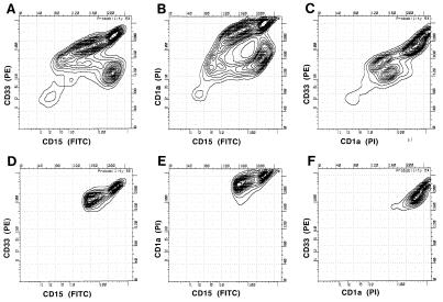
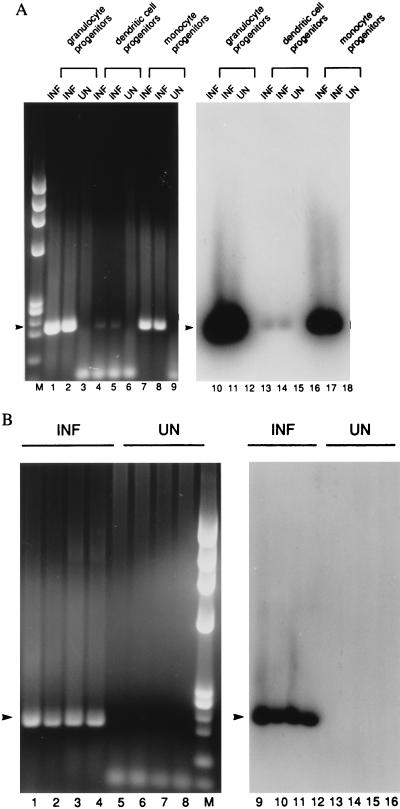
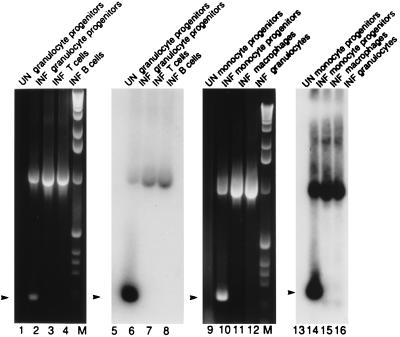
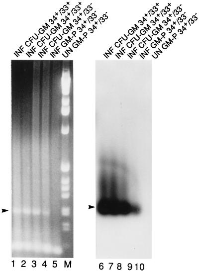
Hematopoietic cells and their progenitors play important roles in human cytomegalovirus latency and reactivation. Latent infection has been evaluated in defined populations of myeloid-lineage-committed progenitor cells coexpressing CD33 and CD15 or CD33 and CD14 along with the dendritic cell markers CD1a and CD10. These CD33+ cell populations were found to support latency and expression of viral latency-associated transcripts and to undergo reactivation of productive viral replication when differentiated in the presence of human fibroblasts. Reactivation was also observed when myeloid cells were carried in the presence of fibroblast-conditioned medium or medium supplemented with certain cytokines (interferon gamma, tumor necrosis factor alpha, interleukin 4, or granulocyte-macrophage colony-simulating factor), suggesting that cell differentiation pathways act as determinants of reactivation. More primitive CD34+ hematopoietic cells were also found to be susceptible to viral infection and latency was maintained as these cells differentiated into CD33+-lineage-committed populations. Between 0.01% and 0.001% of CD33+ CD14+ or CD33+ CD15+ bone marrow mononuclear cells isolated from naturally infected individuals were found to express latent transcripts. Thus, cytomegalovirus is carried within a small percentage of myeloid and dendritic cell progenitors in the healthy seropositive host. Virus reactivation may be triggered by factors associated with the inflammatory response.


Figures

Figure 1
Figure 1
Flow cytometry analysis of infected CD33+ CD15+ CD1a+ dendritic lineage cells. Three-color analysis of CMV-infected progenitors 3 weeks p.i. was performed with anti-CD33 (PE), anti-CD1a (TC, read on the propidium iodide channel), and anti-CD15 (FITC). A–C show the FACS profile of analyzed cells. DF show the sorted populations.
Figure 2
Figure 2
RT-PCR analysis to detect latent transcripts in pools of 300 FACS-sorted monocyte-, granulocyte-, and dendritic-lineage cells 3 weeks p.i. RT-PCR for sense CLTs yields a 206-bp product (arrowhead). (A) The following cell types were analyzed. Lanes: 1–3 and 9–11, granulocyte progenitors (CD33+ CD15+); 4–6 and 12–15, dendritic cell progenitors (CD33+ CD10+ CD15+); 7–9 and 16–18, monocyte progenitors (CD33+ CD14+). (B) The following dendritic cell progenitors were analyzed. Lanes: 1, 2, 5, 6, 9, 10, 13, and 14, CD33+ CD1a+ CD15+; 3, 4, 7, 8, 11, 12, 15, and 16, CD33+ CD1a+ CD14+. (Left) Ethidium bromide-stained gel. (Right) DNA blot. INF, infected cells; UN, uninfected cells; M, molecular weight marker HaeIII-digested φX174.
Figure 3
Figure 3
RT-PCR analysis to detect latent transcripts in 300 infected and sorted PB mononuclear cells. Cells with the following cell surface phenotypes were analyzed. Lanes: 1, 2, 5, and 6, granulocyte progenitors (CD33+ CD15+); 3 and 7, T cells (CD3+ CD4+); 4 and 8, B cells (CD19+); 9, 10, 13, and 14, monocyte progenitors (CD33int CD14+); 11 and 15, monocyte/macrophages (CD33 CD14+); 12 and 16, granulocytes (CD33 CD15+). Lanes 1–4 and 9–12 show ethidium bromide stained gels, and lanes 5–8 and 13–16 show the DNA blot. Conditions and abbreviations are as in Fig. 2. M, molecular weight marker 1-kb ladder. Arrowhead, sense CLTs (206 bp).
Figure 4
Figure 4
RT-PCR analysis to detect latent transcripts in CFU-GM colonies picked from methylcellulose culture. CFU-GM colonies originated from the following FACS-sorted and infected populations. Lanes: 1, 2, 6, and 7, CD34+ CD33+ cells; 3 and 8, CD34+ CD33 cells; 4, 5, 9, and 10, CD34+ CD33 cells grown in liquid culture for 3 weeks and then sorted into CD33+ CD15+ cells. (Left) Ethidium bromide-stained gel. (Right) DNA blot. Conditions and abbreviations are as in Fig. 2. M, molecular weight marker 1-kb ladder.

References

    1. Alford C A, Britt W J. In: Fields Virology. Fields B N, Knipe D M, Howley P M, editors. New York: Lippincott-Raven; 1996. pp. 2493–2534.
    1. Ibanez C E, Schrier R, Ghazal P, Wiley C, Nelson J A. J Virol. 1991;65:6581–6588. - PMC - PubMed
    1. Lathey J L, Spector S A. J Virol. 1991;65:6371–6375. - PMC - PubMed
    1. Maciejewski J P, Bruening E E, Donahue R E, Sellers S E, Carter C, Young N S, St. Jeor S. Virology. 1993;195:327–336. - PubMed
    1. Minton E J, Tysoe C, Sinclair J H, Sissons J G. J Virol. 1994;68:4017–4021. - PMC - PubMed

Publication types