. 1998 Mar 31;95(7):3996-4001.
doi: 10.1073/pnas.95.7.3996.

A tripotential glial precursor cell is present in the developing spinal cord

Affiliations

A tripotential glial precursor cell is present in the developing spinal cord

M S Rao et al. Proc Natl Acad Sci U S A. .

Abstract

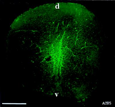
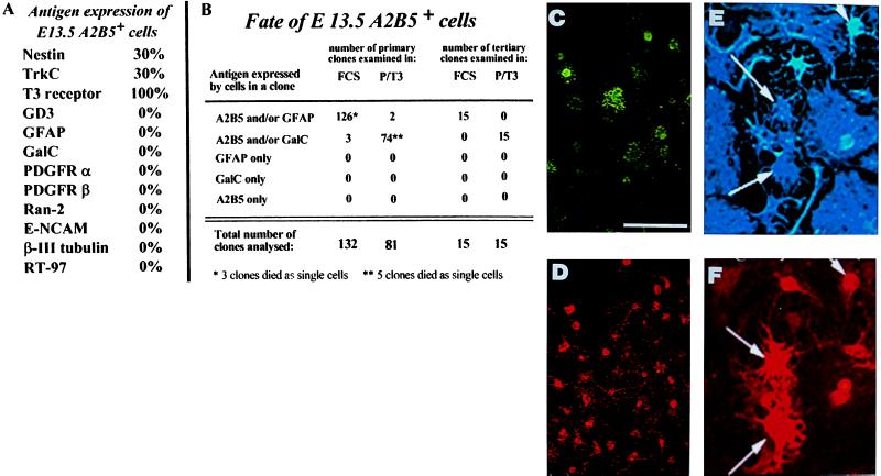
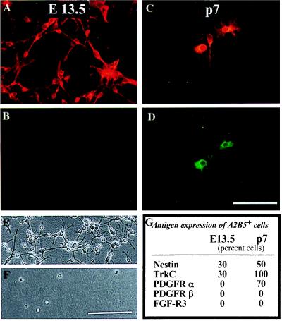
We have isolated a tripotential glial precursor cell population from spinal cords of E13.5 rats. In vitro, these A2B5+E-NCAM- glial-restricted precursor (GRP) cells can undergo extensive self-renewal, and can differentiate into oligodendrocytes and two distinct astrocyte populations, but do not differentiate into neurons. The differentiation potential of GRP cells is retained through at least three cycles of expansion and recloning. Unlike oligodendrocyte-type 2 astrocyte progenitor cells, freshly isolated GRP cells do not respond to platelet-derived growth factor as a mitogen or survival factor, nor do GRP cells differentiate into oligodendrocytes--or even survive--when plated in mitogen-free chemically defined medium. Exposure to fetal calf serum induces GRP cells to differentiate into A2B5- fibroblast-like astrocytes, whereas growth in the presence of basic fibroblast growth factor and ciliary neurotrophic factor induces the generation of A2B5+ process-bearing astrocytes. The early appearance of GRP cells during spinal cord development suggests that they may represent the earliest GRP cell population.


Figures

Figure 1
Figure 1
A2B5+ cells can be detected in E13.5 spinal cord. Spinal cords from E13.5 spinal cords were fresh frozen and sectioned on a cryostat to obtain 10-μm sections. Sections were incubated with A2B5 antibody for 5 hr, washed, and incubated with fluorescein-conjugated goat anti-mouse IgM secondary antibody. A2B5+ cells were seen predominantly in the central region of the spinal cord. (Bar = 140 μm).
Figure 2
Figure 2
A2B5+ cells derived from E13.5 spinal cord are multipotent GRP cells. A2B5+ cells were immunopurified from E13.5 rat spinal cord and grown as clones in medium supplemented with PDGF + bFGF. (A) A2B5+ cells did not express markers of neuroblasts, neurons, astrocytes, or oligodendrocytes. (B) After 5 days of growth in PDGF + bFGF, cloning dishes were switched to medium containing either FCS or PDGF + T3 for 5 days. In dishes of primary clones switched to FCS, all clones generated A2B5+ and A2B5 astrocytes. Clones grown in PDGF + T3 contained oligodendrocytes and GRP cells but few astrocytes. The total of 132 clones in FCS and 81 clones in PDGF + T3 represents the sum of three independent experiments. To examine whether GRP cells were capable of extensive self-renewal without loss of differentation potential, five primary clones were recloned in PDGF + bFGF. Three to five clones were selected randomly from each set of secondary clones and recloned a third time. Dishes were switched to medium containing either FCS or PDGF + T3, and in each condition, three descendant clones of each initial clone were chosen for analysis thus yielding the 15 clones analyzed in each condition. As shown, the ability of the freshly cloned GRPs to differentiate into oligodendrocytes and two types of astrocytes was retained fully, even after tertiary cloning. (CF) A single primary A2B5+ clone that was recloned into separate dishes and induced to differentiate for 5 days in the presence of PDGF + T3 (C and D) or FCS (E and F), after which cultures were immunolabeled. A2B5 staining (D and F) is red, anti-GalC staining (C) is green, and anti-GFAP staining (E) is blue. A clone derived from a single A2B5 cell can differentiate into A2B5+GFAP+ astrocytes (arrows in E and F), A2B5/GFAP+ astrocytes (unlabeled nonprocess-bearing astrocytes in E, and A2B5 GalC+ oligodendrocytes (C). This recloning experiment was repeated three times, from three independent dissections, with identical results. (Bar = 25 μm for C and D, 50 μm for E and F).
Figure 3
Figure 3
GRP cells exhibit extensive self-renewal potential. A2B5+ GRP cells were immunopurified from E13.5 spinal cord and expanded in PDGF + bFGF. Each of the five randomly selected primary clones gave rise to from 66 to 260 secondary clones, and each of the randomly selected secondary clones gave rise to 2–132 tertiary clones. An example of an expanded clone is shown as a phase image and labeled as living cells with A2B5 antibody. (Bar = 40 μm for days 1 and 3; 400 μm for day 10).
Figure 4
Figure 4
GRP cells and O-2A progenitor cells have distinct phenotypes. A2B5+ cells were immunopurified from E13.5 spinal cord or P7 corpus callosum, allowed to adhere overnight, and stained with indicated antibodies (in these experiments, corpus callosum-derived O-2A progenitor cells behave identically to those derived from optic nerve). Double labeling with A2B5 (A and C) and anti-PDGF receptor-α antibodies (B and D) demonstrated that GRPs (A and B), in contrast with O-2A progenitor cells (C and D), did not express detectable levels of this PDGF receptor isoform. In agreement with the receptor expression data, PDGF did not promote survival of GRP cells (F) but, as seen previously (8, 10, 42), was an effective promoter of survival and division of O-2A progenitor cells (E). Other markers that differentiate between these populations are summarized in G; E 13.5 = GRP cells, p7 = O-2A progenitor cells. (Bar = 50 μm for AD; 70 μm day E and F).
Figure 5
Figure 5
GRP cells and O-2A progenitor cells do not respond in the same manner to differentiation signals. Immunopurified GRP cells and O-2A progenitor cells were grown in medium supplemented with either FCS or bFGF + CNTF for 5 or 10 days and then labeled with A2B5 (red), anti-GFAP (green), and anti-GalC (blue) antibodies. GRP cells exposed to bFGF + CNTF differentiated almost entirely into A2B5+ process-bearing astrocytes, and those cells exposed to FCS differentiated almost entirely into flattened A2B5 astrocytes with a fibroblast-like morphology. Oligodendrocytes were seen only rarely in either of these conditions. In contrast, O-2A progenitor cell cultures exposed to bFGF + CNTF contained progenitor cells, oligodendrocytes, and rare type 2 astrocytes (the panel chosen was selected specifically as all three cell types were present in a single field of view but underrepresents the ratio of oligodendrocytes to type 2 astrocytes; see also ref. 25), whereas cultures exposed to FCS showed extensive differentiation into type 2 astrocytes (32, 46). The experiments were repeated three times with identical results. (Bar = 50 μm.)
Figure 6
Figure 6
A2B5+ and A2B5 astrocytes generated from GRP cells differ from each other in expression of the R3 isoform of the FGF receptor and in labeling with the Ran-2 mAB. The A2B5 astrocytes generated from GRP cells in the presence of FCS were predominantly Ran-2+ and expressed FGFR3, whereas the A2B5+ astrocytes generated in the presence of bFGF + CNTF were predominantly Ran-2 and FGFR3. The panel shows labeling of astrocytes, derived from GRP cells grown in the presence of FCS, with antibodies to FGFR3. (Bar = 40 μm.)
Figure 7
Figure 7
Possible relationships between NEP cells, GRP cells and other glial precursor cells. (A) NEP cells may give rise directly to different glial precursor cells of which GRP cells are only one example. (B) NEP cells may give rise to GRP cells as the first step in the generation of precursor cells committed to the production of glia, and GRP cells may then give rise to descendant precursor cells, which are more limited in their differentiation potential. It is also possible to envisage models combining features of A and B.

References

    1. Price J, Williams B, Grove E. Brain Pathol. 1992;2:23–29. - PubMed
    1. Sanes J R. Trends Neurosci. 1989;12:21–28. - PubMed
    1. Goldman J E. J Neuro-Oncol. 1995;24:61–64. - PubMed
    1. Noble M, Gutoswki N, Bevan K, Engel U, Linskey M, Urenjak J, Bhakoo K, Williams S. Glia. 1995;15:222–230. - PubMed
    1. Raff M C. Science. 1989;243:1450–1455. - PubMed

Publication types

Substances