. 1998 Mar 31;95(7):4013-8.
doi: 10.1073/pnas.95.7.4013.

Nurr1 is essential for the induction of the dopaminergic phenotype and the survival of ventral mesencephalic late dopaminergic precursor neurons

Affiliations

Nurr1 is essential for the induction of the dopaminergic phenotype and the survival of ventral mesencephalic late dopaminergic precursor neurons

O Saucedo-Cardenas et al. Proc Natl Acad Sci U S A. .

Abstract

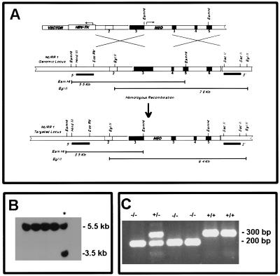
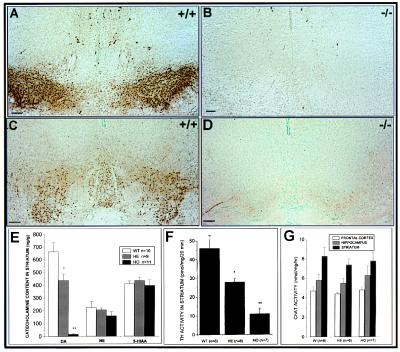
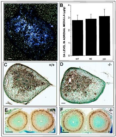
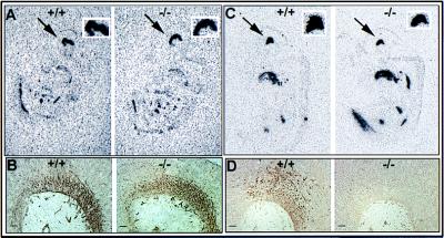
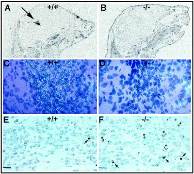
Nurr1 is a member of the nuclear receptor superfamily of transcription factors that is expressed predominantly in the central nervous system, including developing and mature dopaminergic neurons. Recent studies have demonstrated that Nurr1 is essential for the induction of phenotypic markers of ventral mid-brain dopaminergic neurons whose generation is specified by the floor plate-derived morphogenic signal sonic hedgehog (SHH), but the precise role of Nurr1 in this differentiative pathway has not been established. To provide further insights into the role of Nurr1 in the final differentiation pathway, we have examined the fate of dopamine cell precursors in Nurr1 null mutant mice. Here we demonstrate that Nurr1 functions at the later stages of dopamine cell development to drive differentiation of ventral mesencephalic late dopaminergic precursor neurons. In the absence of Nurr1, neuroepithelial cells that give rise to dopaminergic neurons adopt a normal ventral localization and neuronal phenotype characterized by expression of the homeodomain transcription factor and mesencephalic marker, Ptx-3, at embryonic day 11.5. However, these late precursors fail to induce a dopaminergic phenotype, indicating that Nurr1 is essential for specifying commitment of mesencephalic precursors to the full dopaminergic phenotype. Further, as development progresses, these mid-brain dopamine precursor cells degenerate in the absence of Nurr1, resulting in loss of Ptx-3 expression and a concomitant increase in apoptosis of ventral midbrain neurons in newborn null mutant mice. Taken together, these data indicate that Nurr1 is essential for both survival and final differentiation of ventral mesencephalic late dopaminergic precursor neurons into a complete dopaminergic phenotype.


Figures

Figure 1
Figure 1
Targeted inactivation of Nurr1 gene in mouse ES cells and generation of mutant mice. (A) Schematic diagram of the strategy used to target Nurr1 gene. (Top) The 13.7-kb targeting vector used for electroporation. Numbered boxes represent exons. The PGK-neobpA and the HSV-thymidine kinase cassettes are indicated by open boxes. The arrows indicate the direction of transcription. (Middle) Genomic structure of the Nurr1 gene. (Bottom) Representation of the structure of the inactivated Nurr1 gene. (B) Southern blot analysis of the DNA from G418-resistant ES cells. The DNA was digested with BamHI and hybridized to a 900-bp genomic probe located upstream of the 5′ homologous arm. This probe hybridized to a 5.5-kb and a 3.5-kb fragment from wild-type and mutant alleles, respectively. (C) PCR analysis of mice derived from heterozygous Nurr1 (+/−) crosses. Two PCR products are shown at 300 bp and 200 bp correspond to the wild-type and mutant alleles, respectively.
Figure 2
Figure 2
Analysis of monoamines and Chat levels in newborn mice. Coronal sections (A and C) show the TH and AADC immunostaining, respectively, in the substantia nigra (A9) and ventral tegmental (A10) area of wild-type brain. (B and D) Loss of both markers at the same level in coronal sections of nurr1−/− mice. (E) Monoamine levels were measured in the striatum of newborn wild-type, heterozygous, and homozygous mice by HPLC. A complete loss of dopamine levels in the homozygous mice and a significant decrease in the heterozygous mice was detected. The TH activity in the striatum (F) was significantly decreased in the homozygous and heterozygous mice, while choline acetyltransferase activity (G), norepinephrine, and serotonin (E) were uneffected. (Bars = 100 μm.)
Figure 3
Figure 3
TH expression and dopamine levels in neural crest-derived cells of the newborn adrenal medulla. (A) In situ hybridization analysis of Nurr1 mRNA expression in the adrenal gland of a wild-type mouse. Nurr1 mRNA was detected in the medulla of the adrenal gland. (B) Dopamine levels in adrenal medulla of wild-type, heterozygous, and homozygous mice. No significant differences in dopamine levels were detected. TH immunoreactivity of wild-type (C) and mutant adrenal gland from newborn mice (D). (E and F) No detectable differences in TH immunoreactivity in the periglomerular dopaminergic cells of the olfactory bulb in wild-type and nurr1 null mutant mice, respectively. (Bar = 50 μm.)
Figure 4
Figure 4
Analysis of the phenotype of cells in the ventral midbrain of wild-type and Nurr1 mutant embryos. (A) Autoradiographic localization of hybridization to the ventral marker, HNF3β. The arrow shows the strong staining in the ventral part of the midbrain of both 12.5 days wild-type (Left) and mutant (Right) mice embryo. The square on the top shows a higher magnification of this expression. (B) Immunostaining for the general neuronal marker, 3A10, in 11.5 day wild-type and mutant mouse embryos. (C) In situ hybridization analysis of Ptx-3 mRNA expression in 11.5 day mouse embryo. The arrow indicates the positive staining in the ventral midbrain. Wild-type and mutant mice showed similar staining. (D) Immunohistochemical localization of TH expression in the ventral midbrain of 12.5 day wild-type embryo and lack of expression in the nurr1−/− midbrain. (Bars = 20 μm.)
Figure 5
Figure 5
Loss of Ptx-3 expression and increased cell death specifically in the ventral midbrain region of newborn Nurr1−/− mice. (A and C) Localization of Ptx-3 expression in the ventral midbrain (arrow) of wild-type neonate. (B and D) Ptx-3 staining is almost depleted in the Nurr1−/− midbrain. TUNEL staining in the ventral midbrain region of wild-type (E) and Nurr1−/− mice (F). Notice the increase in TUNEL positive nuclei (arrowheads) and dying cells (asterisk) in the Nurr1−/− mice. (Bars = 20 μm.)

References

    1. Bjorklund A, Lindvall O. In: Handbook of Chemical Neuroanatomy. Bjorklund A, Hokfelt T, editors. Amsterdam: Elsevier; 1984. pp. 55–122.
    1. Lindvall O, Bjorlund A. In: Chemical Neuroanatomy. Emson P C, editor. New York: Raven; 1983. pp. 229–255.
    1. Hirsch E C, Graybiel A M, Agid Y A. Nature (London) 1988;334:345–348. - PubMed
    1. Self D W, Nestler E J. Annu Rev Neurosci. 1995;18:463–495. - PubMed
    1. Seeman P, Guan H C, Van Tol H H M. Nature (London) 1997;365:441–445. - PubMed

Publication types