. 1998 Apr;72(4):2647-54.
doi: 10.1128/JVI.72.4.2647-2654.1998.

Generation of a mutant infectious bursal disease virus that does not cause bursal lesions

Affiliations

Generation of a mutant infectious bursal disease virus that does not cause bursal lesions

K Yao et al. J Virol. 1998 Apr.

Abstract

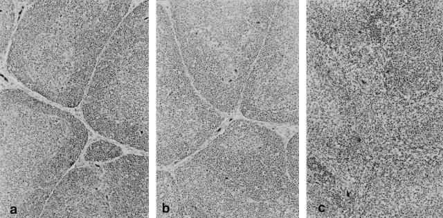
A reverse genetics system for birnavirus, based on synthetic transcripts of the infectious bursal disease virus (IBDV) genome, was recently developed (E. Mundt and V. N. Vakharia, Proc. Natl. Acad. Sci. USA 93:11131-11136, 1996). To study the function of the 17-kDa nonstructural (NS) protein in viral growth and pathogenesis, we constructed a cDNA clone of IBDV segment A in which the first and only initiation codon (ATG) of NS protein was mutated to a stop codon (TAG). Transfection of Vero cells with combined transcripts of either modified or unmodified segment A, and with segment B, generated viable IBDV progeny. When chicken embryo fibroblast cells infected with transfectant viruses were analyzed by immunofluorescence assays using NS-specific antiserum, the mutant virus did not yield a fluorescence signal, indicating a lack of NS protein expression. Furthermore, replication kinetics and cytotoxic effects of the mutant virus were compared with those of the parental attenuated vaccine strain of IBDV (D78) in vitro. The mutant virus grew to slightly lower titers than D78 virus and exhibited decreased cytotoxic and apoptotic effects in cell culture. To evaluate the characteristics of the recovered viruses in vivo, we inoculated 3-week-old chickens with D78 or mutant virus and analyzed their bursa for histopathological lesions. The recovered D78 virus caused microscopic lesions and atrophy of the bursa, while the mutant virus failed to induce any pathological lesions or clinical signs of disease. In both instances, the virus was recovered from the bursa, and the presence or absence of mutation in these viruses was confirmed by nucleotide sequence analysis of NS gene. Although the mutant virus exhibited a delay in replication in vivo, it induced levels of IBDV neutralizing antibodies that were similar to those of D78 virus. In addition, no reversion of mutation was detected in the mutant virus recovered from inoculated chickens. These results demonstrate that NS protein is dispensable for viral replication in vitro and in vivo and that it plays an important role in viral pathogenesis. Thus, generation of such NS protein-deficient virus will facilitate the study of immunosuppression and aid in the development of live-attenuated vaccines for IBDV.


Figures

FIG. 1
FIG. 1
Schematic presentation of IBDV cDNA constructs for the generation of plus-sense RNA transcripts with T7 RNA polymerase. Plasmid pUC19FLAD78 encodes the polyprotein (VP2-VP4-VP3) and the NS protein (NS) of D78-IBDV. In plasmid pUCD78NSΔ, the initiation codon of NS gene is mutated to a stop codon. Plasmid pUCD78B encodes the RNA-dependent RNA polymerase (VP1). All plasmids contain a T7 promoter sequence at their 5′-ends. Plasmids were linearized with the appropriate restriction enzymes as indicated (↑).
FIG. 2
FIG. 2
Immunofluorescence staining of IBDV-infected cells used to detect NS protein expression. CEF cells were infected with passage 1 (b) or passage 10 (e) rD78NSΔ mutant virus stock or with passage 1 (c) or passage 10 (f) rD78 virus stock at an MOI of 1. Uninfected CEF cells were used as negative controls (a and d). After 24 h postinfection, the cells were fixed and analyzed by immunofluorescence staining with rabbit anti-NS protein serum. Magnifications, ×400.
FIG. 3
FIG. 3
Growth curve of IBDV (A) and cytotoxic effect in IBDV-infected cells (B). (A) Monolayers of CEF cells were infected with the indicated viruses at an MOI of 0.1 and harvested at the indicated time points, and infectious titers were determined by plaque assay. (B) Cell cultures were assayed for viability at the indicated times by the MTT assay. The mean of the OD value ≥1.0 unit above that of control wells at each day was defined as 100% cell survival. Each value is the average of two independent experiments.
FIG. 4
FIG. 4
Analysis of IBDV-induced apoptosis by TUNEL assay. CEF cells were either mock infected (a, d, and g) or infected with rD78NSΔ (b, e, and h) or rD78 (c, f, and i) IBDV at an MOI of 1. Cells were fixed at 24 h (a to c), 48 h (d to f), and 72 h (g to i) postinfection and assayed by immunofluorescence for DNA breakage (TdT assay; green signal). The TUNEL staining of CEF cells, infected with NS protein-deficient mutant IBDV, show a substantial reduction in IBDV-induced apoptosis. Magnifications, ×100.
FIG. 5
FIG. 5
Histopathologic appearance of sections (hematoxylin and eosin) of BF derived from mock-infected and infected chickens at day 6 postinfection. (a) Cortical lymphocytes (dark gray cells adjacent to connective tissue that separates follicles) and medullary lymphocytes (light gray cells in follicle centers) in portions of six follicles from an uninfected chicken are normal. In addition, the interfollicular connective tissues are normal. (b) Follicles and interfollicular connective tissues from a chicken infected with the mutant rD78NSΔ virus are normal and cannot be differentiated from their control counterparts. (c) There is lymphocytic necrosis and heterophilic inflammation in six follicles in the BF from a chicken infected with rD78 virus. Notice the loss of distinction between the cortex and the medulla and the bands of interfollicular connective tissue that are infiltrated by myriad heterophils and macrophages. Magnifications, ×100.

References

    1. Ball J M, Tian P, Zeng C Q-Y, Morris A P, Estes M K. Age-dependent diarrhea induced by a rotaviral nonstructural glycoprotein. Science. 1996;272:101–104. - PubMed
    1. Böttcher B, Kiselev N A, Stel’Mashchuk V Y, Perevozchikova N A, Borisov A V, Crowther R A. Three-dimensional structure of infectious bursal disease virus determined by electron microscopy. J Virol. 1997;71:325–330. - PMC - PubMed
    1. Brown C C, Piccone M E, Mason P W, McKenna T S-C, Grubman M J. Pathogenesis of wild-type and leaderless foot-and-mouth disease virus in cattle. J Virol. 1996;70:5638–5641. - PMC - PubMed
    1. Dobos P, Hill B J, Hallet R, Kells D T C, Becht H, Teninges D. Biophysical and biochemical characterization of five animal viruses with bisegmented double-stranded genomes. J Virol. 1979;32:593–605. - PMC - PubMed
    1. Hirai K, Calnek B W. In vitro replication of infectious bursal disease virus in established cell lines and chicken B lymphocytes. Infect Immun. 1979;25:964–970. - PMC - PubMed

Publication types

Substances

LinkOut - more resources