. 1998 Apr;72(4):2846-54.
doi: 10.1128/JVI.72.4.2846-2854.1998.

Sequential steps in human immunodeficiency virus particle maturation revealed by alterations of individual Gag polyprotein cleavage sites

Affiliations

Sequential steps in human immunodeficiency virus particle maturation revealed by alterations of individual Gag polyprotein cleavage sites

K Wiegers et al. J Virol. 1998 Apr.

Abstract

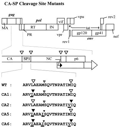
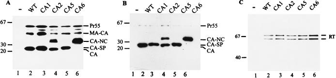
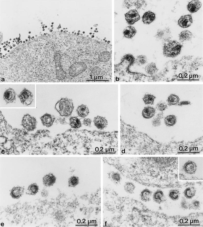
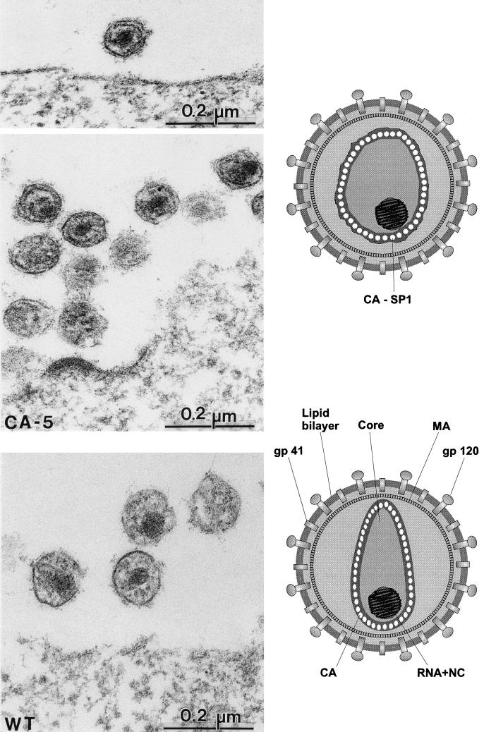
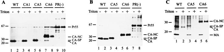
Retroviruses are produced as immature particles containing structural polyproteins, which are subsequently cleaved by the viral proteinase (PR). Extracellular maturation leads to condensation of the spherical core to a capsid shell formed by the capsid (CA) protein, which encases the genomic RNA complexed with nucleocapsid (NC) proteins. CA and NC are separated by a short spacer peptide (spacer peptide 1 [SP1]) on the human immunodeficiency virus type 1 (HIV-1) Gag polyprotein and released by sequential PR-mediated cleavages. To assess the role of individual cleavages in maturation, we constructed point mutations abolishing cleavage at these sites, either alone or in combination. When all three sites between CA and NC were mutated, immature particles containing stable CA-NC were observed, with no apparent effect on other cleavages. Delayed maturation with irregular morphology of the ribonucleoprotein core was observed when cleavage of SP1 from NC was prevented. Blocking the release of SP1 from CA, on the other hand, yielded normal condensation of the ribonucleoprotein core but prevented capsid condensation. A thin, electron-dense layer near the viral membrane was observed in this case, and mutant capsids were significantly less stable against detergent treatment than wild-type HIV-1. We suggest that HIV maturation is a sequential process controlled by the rate of cleavage at individual sites. Initial rapid cleavage at the C terminus of SP1 releases the RNA-binding NC protein and leads to condensation of the ribonucleoprotein core. Subsequently, CA is separated from the membrane by cleavage between the matrix protein and CA, and late release of SP1 from CA is required for capsid condensation.


Figures

FIG. 1
FIG. 1
Schematic representation of various mutants. At the top, the coding region of the HIV-1 genome is shown, with the different open reading frames depicted as boxes. The 3′-terminal part of the CA coding region and the NC coding region of the gag reading frame, including SP1, are expanded in the middle. Cleavage sites for HIV-1 PR are indicated as open triangles. The signal for translational frameshifting is also marked (black arrow). At the bottom, the amino acid sequence of wild-type (WT) SP1 and flanking cleavage sites as well as the altered sequences of the various mutant constructs are shown. The name of each construct is indicated on the left. Mutated amino acids are underlined. Large open triangles indicate cleavage sites that are processed by HIV-1 PR; large closed triangles represent sequences that are not cleaved by HIV-1 PR. The smaller triangles correspond to a cryptic PR cleavage site within SP1 which is cleaved much more slowly.
FIG. 2
FIG. 2
Western blot analysis of gag and pol gene products after transient transfection. COS-7 cells transfected with pNL4-3 or derivatives and media were harvested 72 h after transfection. Lysates of transfected cells (A) or viral particles collected by centrifugation through a sucrose cushion (B and C) were resolved by SDS-polyacrylamide gel electrophoresis, and Western blots were reacted with polyclonal antiserum against CA (A and B) or RT (C) and tested with chemiluminescence. The wild-type (WT) and mutant proviral constructs are indicated above each lane. Molecular mass standards (in kilodaltons) are indicated on the left. HIV-specific precursor proteins, intermediate processing products, and cleaved proteins are indicated on the right.
FIG. 3
FIG. 3
Electron micrographs showing thin sections of COS-7 cells transfected with pNL4-3 and mutant derivatives at 72 h after transfection. (a and b) Overview (a) and higher-magnification view (b) of wild-type-transfected cells and extracellular particles. (c and inset) Mutant pNL43-CA1. (d) Mutant pNL43-CA5. (e) Mutant pNL43-CA2. (f and inset) Mutant pNL43-CA6.
FIG. 4
FIG. 4
Comparative analysis of particle morphology of HIV-1 wild-type (WT) and mutant CA5 particles. Electron micrographs of WT particles show an electron-dense centrally located nucleocapsid surrounded by a conical capsid shell (best seen in the particle at the left). For the CA5 mutant, particles are of a similar size and also contain a centrally located electron-dense nucleocapsid but lack the conical capsid structure. Instead, a thin electron-dense layer is observed in the vicinity of the viral membrane but clearly separated from it. Schematic representations of the morphologies of the different particles are shown on the right.
FIG. 5
FIG. 5
Analysis of detergent stability of wild-type and mutant HIV particles. (A) Virus particles collected from the medium of transfected COS-7 cells were treated with 0.5% Triton X-100 for 10 min or left untreated, and both samples were subsequently centrifuged through a sucrose cushion. (B and C) In a second experiment, virus particles were layered on a sucrose step gradient containing a layer of 10% sucrose with or without 0.5% Triton X-100 on top of a 20% sucrose layer as described in Materials and Methods. Pellet fractions were resolved by SDS-polyacrylamide gel electrophoresis followed by silver staining (C) or Western blot analysis with polyclonal antiserum against CA and chemiluminescence detection (A and B). The wild-type (WT) proviral construct and the respective mutants are indicated above each lane. The presence or absence of 0.5% Triton X-100 is indicated by + or −, respectively. Molecular mass standards (in kilodaltons) are indicated on the left. HIV-specific precursor proteins and cleavage products are identified on the right.

References

    1. Adachi A, Gendelman H E, Koenig S, Folks T, Willey R, Rabson A, Martin M A. Production of acquired immunodeficiency syndrome-associated retrovirus in human and nonhuman cells transfected with an infectious molecular clone. J Virol. 1986;59:284–291. - PMC - PubMed
    1. Berkowitz R, Fisher J, Goff S P. RNA packaging. Curr Top Microbiol Immunol. 1996;214:177–218. - PubMed
    1. Campbell S, Vogt V M. Self-assembly in vitro of purified CA-NC proteins from Rous sarcoma virus and human immunodeficiency virus type 1. J Virol. 1995;69:6487–6497. - PMC - PubMed
    1. Campbell S, Vogt V M. In vitro assembly of virus-like particles with Rous sarcoma virus Gag deletion mutants: identification of the p10 domain as a morphological determinant in the formation of spherical particles. J Virol. 1997;71:4425–4435. - PMC - PubMed
    1. Chen C, Okayama H. High-efficiency transformation of mammalian cells by plasmid DNA. Mol Cell Biol. 1987;7:2745–2752. - PMC - PubMed

Publication types

MeSH terms