. 1998 Apr;72(4):2896-904.
doi: 10.1128/JVI.72.4.2896-2904.1998.

Adenovirus preterminal protein binds to the CAD enzyme at active sites of viral DNA replication on the nuclear matrix

Affiliations

Adenovirus preterminal protein binds to the CAD enzyme at active sites of viral DNA replication on the nuclear matrix

P C Angeletti et al. J Virol. 1998 Apr.

Abstract

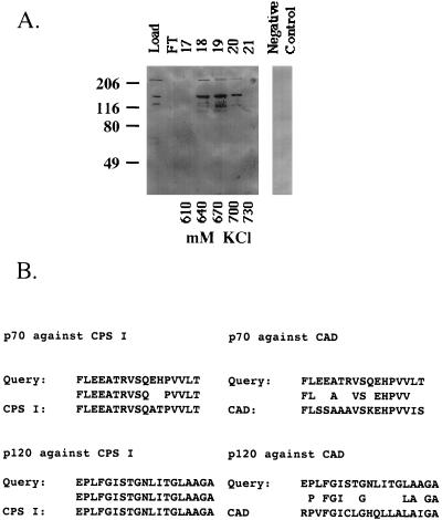
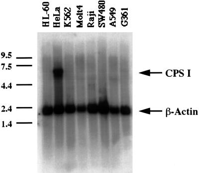
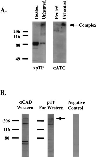
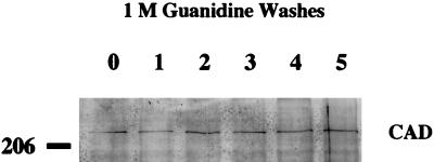
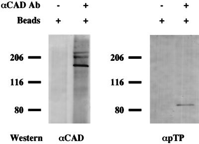
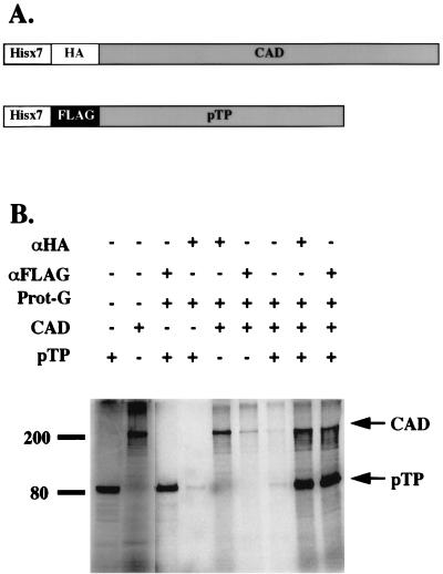
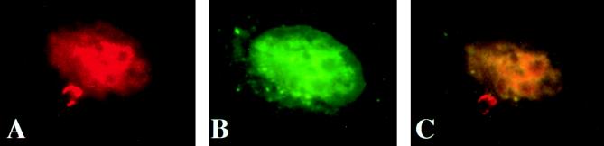
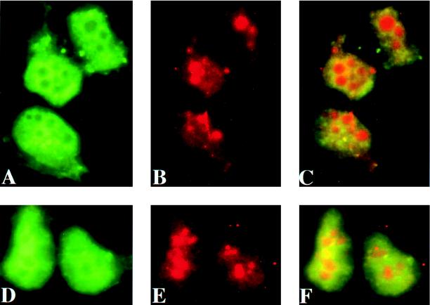
Adenovirus (Ad) replicative complexes form at discrete sites on the nuclear matrix (NM) via an interaction mediated by the precursor of the terminal protein (pTP). The identities of cellular proteins involved in these complexes have remained obscure. We present evidence that pTP binds to a multifunctional pyrimidine biosynthesis enzyme found at replication domains on the NM. Far-Western blotting identified proteins of 150 and 240 kDa that had pTP binding activity. Amino acid sequencing of the 150-kDa band revealed sequence identity to carbamyl phosphate synthetase I (CPS I) and a high degree of homology to the related trifunctional enzyme known as CAD (for carbamyl phosphate synthetase, aspartate transcarbamylase, and dihydroorotase). Western blotting with an antibody directed against CAD detected a 240-kDa band that comigrated with that detected by pTP far-Western blotting. Binding experiments showed that a pTP-CAD complex was immunoprecipitable from cell extracts in which pTP was expressed by a vaccinia virus recombinant. Additionally, in vitro-translated epitope-tagged pTP and CAD were immunoprecipitable as a complex, indicating the occurrence of a protein-protein interaction. Confocal fluorescence microscopy of Ad-infected NM showed that pTP and CAD colocalized in nuclear foci. Both pTP and CAD were confirmed to colocalize with active sites of replication detected by bromodeoxyuridine incorporation. These data support the concept that the pTP-CAD interaction may allow anchorage of Ad replication complexes in the proximity of required cellular factors and may help to segregate replicated and unreplicated viral DNA.


Figures

FIG. 1
FIG. 1
Fractionation of pTP binding proteins and amino acid sequencing. (A) Eleven milligrams of NME was loaded onto a 5-ml P11 phosphocellulose column. The retained protein was eluted with a linear KCl gradient of 100 to 1,000 mM. Twenty-microgram samples representing loaded NME (Load), flowthrough (FT), or eluted fractions (17 to 21) were separated on an 8% polyacrylamide gel and transferred to nitrocellulose. The blot was then probed with a pTP cytoplasmic extract prepared from cells infected with a vaccinia virus recombinant expressing pTP (vvpTP1). An identical blot of the loaded NME was probed with a T7-vaccinia virus recombinant cytoplasmic extract (Negative Control). Two major bands, with molecular masses of 240 and 150 kDa, were detected with pTP. The positions of molecular mass markers are shown to the left (in kilodaltons). (B) Fractions 18 and 19 were pooled, electrophoresed in batch, and blotted to a polyvinylidene difluoride membrane. The pTP-reactive band of 150 kDa was excised and subjected to amino acid sequencing. The amino acid sequences of two peptides (p70 and p120) obtained by endoproteinase Lys-C digestion of the 150-kDa band were determined. The pileup of sequences is the result of a Blast search of the GenBank database. Peptide p70 corresponded to amino acids 1151 to 1167 of CPS I, and p120 corresponded to amino acids 288 to 306 (left). The Blast search also revealed the homology of p70 and p120 to the related CAD enzyme. Peptides p70 and p120 corresponded to amino acid positions 1110 to 1126 and 246 to 264, respectively, of CAD (right).
FIG. 2
FIG. 2
CPS I is expressed only in HeLa cells. A multitumor Northern blot containing 2 μg of poly(A)+ mRNA per lane was probed with a 0.6-kb fragment of the CPS I gene (nucleotides 3471 to 4077). The blot was hybridized at 42°C for 12 h. The lanes contained the following samples: promyelocytic leukemia (HL-60), HeLa cell, mylogenous leukemia (K562), lymphoblastic leukemia (Molt4), Burkitt’s lymphoma (Raji), colorectal carcinoma (SW480), lung carcinoma (A549), and melanoma (G361). CPS I (6.0 kb) is indicated by the upper arrow. The 2.0-kb (β-actin) internal control is indicated by the lower arrow. The positions of molecular size markers are shown to the left (in kilobases).
FIG. 3
FIG. 3
The CAD enzyme comigrates with the 240-kDa NM band detected by pTP far-Western blotting. (A) Thirty micrograms of supernatant from rATP-treated pTP NM was either heated or not in the presence of Laemmli buffer with 0.1% SDS. Samples were applied to an SDS–8% polyacrylamide gel in duplicate. The resultant halves of the blot were probed with either anti-pTP (αpTP) or anti-CAD (αATC) antibodies. The arrow to the right indicates the position of the high-MW pTP- and CAD-containing complex. The positions of molecular mass markers are shown to the left (in kilodaltons). (B) Thirty micrograms of NME was separated on an SDS–8% polyacrylamide gel and blotted to nitrocellulose. A portion of the blot was probed with anti-CAD antibody (αCAD Western). The other lanes were probed with either pTP extract (pTP Far Western) or T7-vaccinia virus cytoplasmic extract as a control for the far-Western blotting (Negative Control). Far-western blot lanes were then developed to show the presence of a pTP interaction with the polyclonal anti-pTP antibody (3-1A). The arrow indicates the 240-kDa band detected by pTP which comigrates with CAD. The positions of molecular mass markers are shown to the left (in kilodaltons).
FIG. 4
FIG. 4
CAD is tightly associated with the NM. Each lane contains 20 μg of uninfected-HeLa-cell NM which was washed with 1 M GnHCl for the number of times indicated above the blot. The blotted protein was probed with anti-CAD antibody raised against the ATC domain. On the left is indicated the position of the 206-kDa molecular mass marker.
FIG. 5
FIG. 5
CAD and pTP coimmunoprecipitate from HeLa cell extracts. Fifty micrograms of whole-cell extract prepared from HeLa cells programmed to express pTP was mixed with either protein A-Sepharose beads alone or beads to which anti-CAD (αCAD) antibody was prebound (indicated by plus and minus symbols). Duplicate immunoprecipitation reaction mixtures were applied to an 8% polyacrylamide gel. Protein blotted to nitrocellulose was probed with either αCAD or antibody to pTP (αpTP). The positions of molecular mass markers are shown to the left of each blot (in kilodaltons).
FIG. 6
FIG. 6
Coimmunoprecipitation of in vitro-translated pTP-CAD fusions. (A) The diagram at the top represents the epitope-tagged constructs that were used. The fusion proteins were cotranslationally labeled with [35S]methionine. (B) The products were mixed with either protein G (Prot-G) alone or protein G prebound with monoclonal antibody to HA (αHA) or FLAG (αFLAG). The combinations for each reaction are indicated above the gel (by plus and minus symbols). The gel was visualized with a Storm PhosphorImager (Molecular Dynamics). Immunoprecipitation of pTP and CAD proteins is indicated by the arrows. The positions of molecular mass standards are shown to the left (in kilodaltons).
FIG. 7
FIG. 7
pTP and CAD colocalize in foci on the NM. HeLa cells were grown on coverslips and infected with Ad for 20 h. The cells were treated with DNase and extracted with 2 M NaCl to create in situ NM. Double staining with anti-pTP monoclonal antibody (IB6A8) and anti-CAD polyclonal antibody specific for the ATC domain was performed. (A) pTP was visualized with TXRD-conjugated goat anti-mouse antibody. (B) CAD was visualized with FITC-conjugated goat anti-rabbit antibody. (C) Merged image of pTP and CAD staining.
FIG. 8
FIG. 8
pTP and CAD colocalize with sites of active replication on the NM. Cells infected with Ad for 20 h were pulsed with BrdU for 20 min. Cells were double stained either with anti-pTP polyclonal antibody (A) and anti-BrdU monoclonal antibody (B) or with anti-CAD polyclonal antibody (D) and anti-BrdU monoclonal antibody (E). Polyclonal antibodies to the CAD ATC domain and pTP were reacted with FITC-conjugated goat anti-rabbit IgG, while the monoclonal antibody to BrdU was detected with TXRD-conjugated goat anti-mouse IgG. (C) Merged image of pTP and BrdU staining. (F) Merged image of CAD and BrdU staining.

Similar articles

Cited by

References

    1. Angeletti P C, Engler J A. Tyrosine kinase-dependent release of an adenovirus preterminal protein complex from the nuclear matrix. J Virol. 1996;70:3060–3067. - PMC - PubMed
    1. Ausubell F M, Brent R, Kingston R E, Moore D D, Seidman J G, Smith J A, Struhl K. Current protocols in molecular biology. New York, N.Y: John Wiley and Sons Inc.; 1995.
    1. Berezney R, Coffey D S. Identification of a nuclear protein matrix. Biochem Biophys Res Commun. 1974;60:1410–1417. - PubMed
    1. Berezney R, Coffey D S. Nuclear matrix: isolation and characterization of a framework structure from rat liver nuclei. J Cell Biol. 1977;73:616–637. - PMC - PubMed
    1. Bidwell J P, Van Wijnen A J, Fey E G, Dworetzky S, Penman S, Stein J L, Lian J B, Stein G S. Osteocalcin gene promoter-binding factors are tissue-specific nuclear matrix components. Proc Natl Acad Sci USA. 1993;90:3162–3166. - PMC - PubMed

Publication types

MeSH terms

LinkOut - more resources