. 1998 Apr;72(4):2935-44.
doi: 10.1128/JVI.72.4.2935-2944.1998.

Functional analysis of the human immunodeficiency virus type 1 Rev protein oligomerization interface

Affiliations

Functional analysis of the human immunodeficiency virus type 1 Rev protein oligomerization interface

S L Thomas et al. J Virol. 1998 Apr.

Abstract

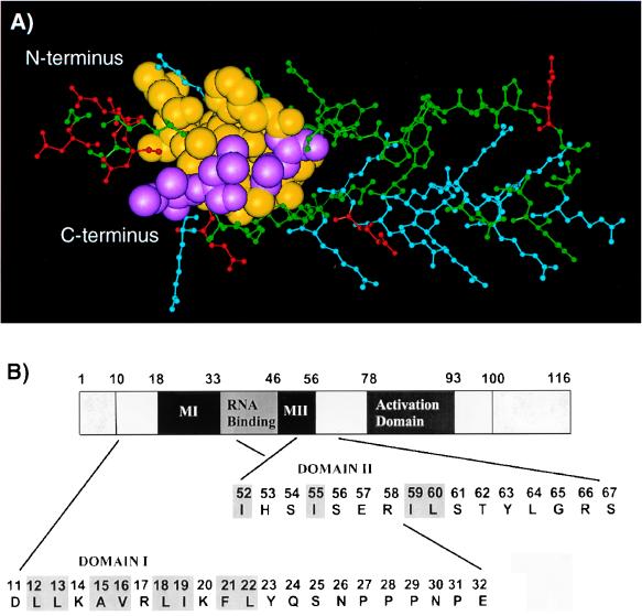
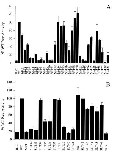
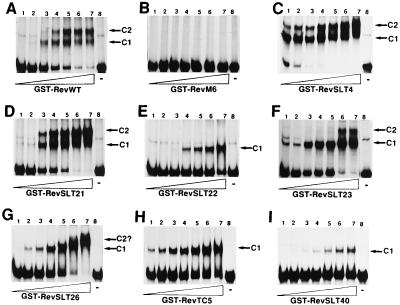
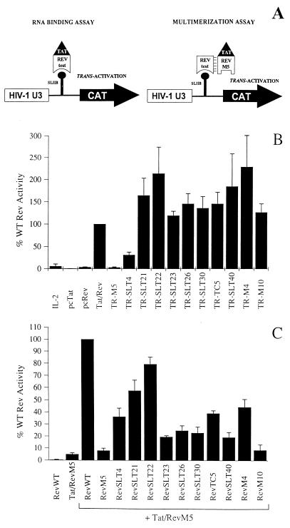
The expression of human immunodeficiency virus type 1 (HIV-1) structural proteins requires the action of the viral trans-regulatory protein Rev. Rev is a nuclear shuttle protein that directly binds to its cis-acting Rev response element (RRE) RNA target sequence. Subsequent oligomerization of Rev monomers on the RRE and interaction of Rev with a cellular cofactor(s) result in the cytoplasmic accumulation of RRE-containing viral mRNAs. Moreover, Rev by itself is exported from the nucleus to the cytoplasm. Although it has been demonstrated that Rev multimerization is critically required for Rev activity and hence for HIV-1 replication, the number of Rev monomers required to form a trans-activation-competent complex on the RRE is unknown. Here we report a systematic analysis of the putative multimerization domains within the Rev trans-activator protein. We identify the amino acid residues which are part of the proposed single hydrophobic surface patch in the Rev amino terminus that mediates intermolecular interactions. Furthermore, we show that the expression of a multimerization-deficient Rev mutant blocks HIV-1 replication in a trans-dominant (dominant-negative) fashion.


Figures

FIG. 1
FIG. 1
Location of the putative HIV-1 Rev multimerization domains. (A) Model of the Rev amino terminus, redrawn from Thomas et al. (100), shows the helix-loop-helix motif of Rev residues 1 to 60. It is evident that the two putative multimerization domains form a single exposed hydrophobic surface patch which presumably constitutes an oligomerization interface. Amino acids are colored as follows: basic residues (Arg, Lys), blue; acidic residues (Asp, Glu), red; residues analyzed in this study, yellow; and residues identified in this study as important for intermolecular interactions, purple. (B) Diagrammatic representation of the 116-aa Rev trans-activator protein showing the functional domains and the regions mutagenized. The two protein regions that form a putative multimerization surface are marked as domains MI and MII. The amino acid sequences of both regions systematically mutagenized in this study are shown as expanded sections. Residues potentially involved in multimerization and previously identified by examination of the refined structural model of Rev (100) are highlighted by shaded boxes.
FIG. 2
FIG. 2
Biological activities of HIV-1 Rev mutant proteins. (A) Scanning mutagenesis of putative multimerization domain MI. Rev trans-activation capacity was determined by cotransfection of COS cell monolayers with the Rev-responsive reporter plasmid pDM128/CMV, the various mutant Rev expression plasmids (indicated on the x axis; see also Table 1), and the constitutive internal control vector pBC12/CMV/βGal. Data are expressed as a percentage of wild-type (WT) Rev activity (set to 100%), and the error bars represent the standard deviations of four independent experiments. All CAT values were adjusted for transfection efficiency by determining the level of β-galactosidase in each culture and were corrected for background (mock) activity. IL-2, negative control plasmid pBC12/CMV/IL-2; WT, wild-type Rev (pcRev). (B) Scanning mutagenesis of putative multimerization domain MII. Details are as for panel A.
FIG. 3
FIG. 3
In vitro RRE binding analysis of wild-type and mutant GST-Rev proteins. A constant level of a 32P-labelled RRE RNA probe was incubated with increasing amounts (30 ng to 2 μg, lanes 1 to 7) of wild-type Rev (RevWT) (A) or the indicated Rev mutant fusion proteins (B to I) and then subjected to RNA gel mobility shift analysis. In each case, lane 8 contained free (unbound) RRE RNA. The various distinct complexes formed upon Rev binding to the RRE RNA probe (C1 and C2) are indicated to the right of each panel.
FIG. 4
FIG. 4
RNA binding and multimerization activities of nonfunctional Rev mutant proteins in a mammalian cell culture system. (A) Diagrammatic representation of the pSLIIB/CAT reporter system. Activation of a chimeric HIV-1 LTR promoter in which the TAR sequence was replaced by the RRE SLIIB element (101) allowed the monitoring of in vivo RRE SLIIB RNA binding and multimer formation by Rev (see the text for further details). (B) In vivo RNA binding activity of nonfunctional Rev mutants. HeLa cells were cotransfected with reporter construct pSLIIB/CAT, the various Tat-Rev (TR) expression plasmids (indicated on the x axis), and constitutive internal control plasmid pBC12/CMV/βGal. pBC12/CMV/IL-2 (IL-2), pcTat, pcRev, and pcTat/Rev (Tat/Rev) were included as controls. At 42 h posttransfection, protein extracts were prepared to assay for β-galactosidase and CAT levels. Data are expressed as a percentage of wild-type (WT) Rev activity (set to 100%) and were corrected for background (mock) activity. (C) Analysis of Rev multimerization in vivo. The ability of the selected nonfunctional Rev mutants to form multimers was determined by cotransfection of HeLa cells with reporter construct pSLIIB/CAT, pcTat/RevM5 (an RNA binding-negative mutant), the various mutant Rev expression plasmids (indicated on the x axis), and constitutive internal control plasmid pBC12/CMV/βGal. pcRev (RevWT) and pcTat/RevM5 (Tat/RevM5) alone were included as negative controls. At 42 h posttransfection, levels of β-galactosidase and CAT were determined as described above. Data are expressed as a percentage of wild-type (WT) Rev activity (set to 100%). Error bars represent the standard deviations of four independent experiments.
FIG. 5
FIG. 5
Competitive inhibition of Rev function. COS cell cultures were cotransfected with the HXB-2-derived Rev-deficient proviral DNA HIV-1Δrev, pBC12/RSV/SEAP (internal control vector), and pBC12/CMV/IL-2 (negative), pcRev (positive), or pcRev plus a 10-fold excess of expression vector encoding wild-type (WT) Rev, RevSLT26, RevSLT40, or RevM10. Total input DNA was maintained at a constant level by inclusion of parental vector pBC12/CMV. All p24 Gag antigen values were adjusted for transfection efficiency by determining the level of SEAP (5) in each culture and were corrected for background (mock) activity. Error bars represent the standard deviations of three independent experiments.

Similar articles

Cited by

References

    1. Auer M, Gremlich H-U, Seifert J-M, Daly T J, Parslow T G, Casari G, Gstach H. Helix-loop-helix motif in HIV-1 Rev. Biochem. 1994;33:2988–2996. - PubMed
    1. Bahner I, Zhou C, Yu X-J, Hao Q-L, Guatelli J C, Kohn D B. Comparison of trans-dominant inhibitory mutant human immunodeficiency virus type 1 genes expressed by retroviral vectors in human T lymphocytes. J Virol. 1993;67:3199–3207. - PMC - PubMed
    1. Bartel D P, Zapp M L, Green M R, Szostak J W. HIV-1 Rev regulation involves recognition of non-Watson-Crick base pairs in viral RNA. Cell. 1991;67:529–536. - PubMed
    1. Berger J, Aepinus C, Dobrovnik M, Fleckenstein B, Hauber J, Böhnlein E. Mutational analysis of functional domains in the HIV-1 Rev trans-regulatory protein. Virology. 1991;183:630–635. - PubMed
    1. Berger J, Hauber J, Hauber R, Geiger R, Cullen B R. Secreted placental alkaline phosphatase: a powerful new quantitative indicator of gene expression in eukaryotic cells. Gene. 1988;66:1–10. - PubMed

Publication types

Substances

LinkOut - more resources